

Function of the chondrocyte PI-3 kinase-Akt signaling pathway is stimulus dependent
- PMID: 25659655
- PMCID: PMC4444401
- DOI: 10.1016/j.joca.2015.01.014
Function of the chondrocyte PI-3 kinase-Akt signaling pathway is stimulus dependent
Abstract
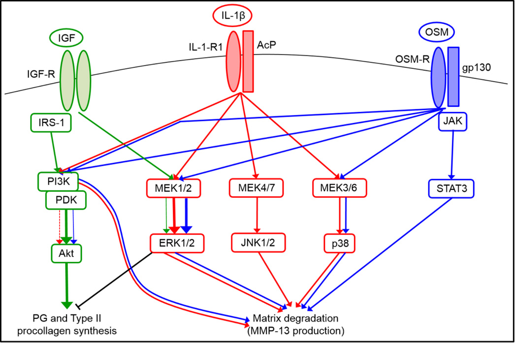
Objective: The PI-3 kinase-Akt pathway plays a role in cartilage anabolic as well as catabolic processes in response to activation by insulin-like growth factor-1 (IGF-1) and the pro-inflammatory cytokines interleukin-1β (IL-1β) and oncostatin M (OSM). The goal of this study was to determine how PI-3 kinase-Akt signaling regulates these seemingly opposing functions.
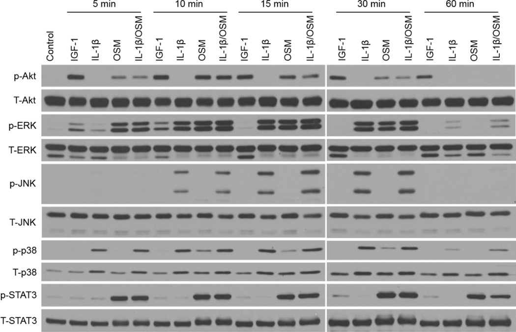
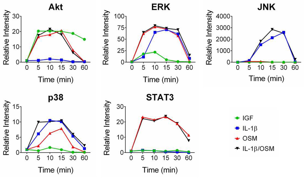
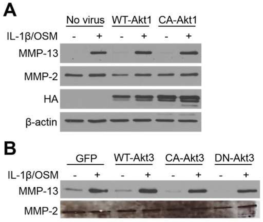
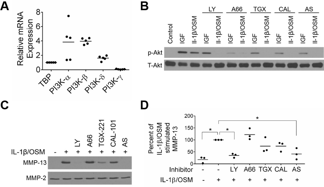
Design: Monolayer cultures of primary human articular chondrocytes were treated with IGF-1, IL-1β, OSM, or the combination of IL-1β and OSM in time course experiments. Activation of signaling proteins and MMP production were measured by immunoblotting. Cells were pre-treated with chemical inhibitors to block mitogen activated protein (MAP) kinases, PI-3 kinase, or JAK/STAT pathway activation. Constitutively active Akt1 and Akt3 were expressed to study stimulus-independent activation of Akt.
Results: IGF-1, OSM, and the combination of IL-1β and OSM but not IL-1β alone, stimulated phosphorylation of Akt which was sustained longer with IGF-1. IL-1β plus OSM, but not IGF-1, increased chondrocyte MMP-13 production which was inhibited with either a general PI-3 kinase inhibitor or specific inhibition of the PI-3 kinase-γ isoform. Akt1 or Akt3 activity alone was not sufficient to increase production of MMP-13. IL-1β/OSM induced MMP-13 production required activation of the MAP kinases, JNK and p38, as well as the JAK-STAT pathway which were activated by IL-1β plus OSM but not by IGF-1.
Conclusions: The chondrocyte integrates signals from the PI-3 kinase-Akt pathway with signals from MAP kinases and the JAK-STAT pathway to allow for a differential response to a pro-anabolic (IGF-1) and a pro-catabolic (IL-1β plus OSM) stimulus.
Keywords: Cell signaling; Chondrocyte; Matrix metalloproteinase-13; PI-3 kinase.
Copyright © 2015 Osteoarthritis Research Society International. Published by Elsevier Ltd. All rights reserved.
Conflict of interest statement
The authors declare no conflicts of interest.
Figures

Similar articles
-
Signaling pathways implicated in oncostatin M-induced aggrecanase-1 and matrix metalloproteinase-13 expression in human articular chondrocytes.Biochim Biophys Acta. 2007 Mar;1773(3):309-20. doi: 10.1016/j.bbamcr.2006.11.018. Epub 2006 Dec 15. Biochim Biophys Acta. 2007. PMID: 17208315
-
Oncostatin M-induced matrix metalloproteinase and tissue inhibitor of metalloproteinase-3 genes expression in chondrocytes requires Janus kinase/STAT signaling pathway.J Immunol. 2001 Mar 1;166(5):3491-8. doi: 10.4049/jimmunol.166.5.3491. J Immunol. 2001. PMID: 11207308
-
IGF-1 and PDGF-bb suppress IL-1β-induced cartilage degradation through down-regulation of NF-κB signaling: involvement of Src/PI-3K/AKT pathway.PLoS One. 2011;6(12):e28663. doi: 10.1371/journal.pone.0028663. Epub 2011 Dec 14. PLoS One. 2011. PMID: 22194879 Free PMC article.
-
Biology and pathology of Rho GTPase, PI-3 kinase-Akt, and MAP kinase signaling pathways in chondrocytes.J Cell Biochem. 2010 Jun 1;110(3):573-80. doi: 10.1002/jcb.22604. J Cell Biochem. 2010. PMID: 20512918 Free PMC article. Review.
-
Multifaceted oncostatin M: novel roles and therapeutic potential of the oncostatin M signaling in rheumatoid arthritis.Front Immunol. 2023 Nov 1;14:1258765. doi: 10.3389/fimmu.2023.1258765. eCollection 2023. Front Immunol. 2023. PMID: 38022540 Free PMC article. Review.
Cited by
-
Chondrocyte Aging: The Molecular Determinants and Therapeutic Opportunities.Front Cell Dev Biol. 2021 Jul 14;9:625497. doi: 10.3389/fcell.2021.625497. eCollection 2021. Front Cell Dev Biol. 2021. PMID: 34336816 Free PMC article. Review.
-
Response eQTLs, chromatin accessibility, and 3D chromatin structure in chondrocytes provide mechanistic insight into osteoarthritis risk.Cell Genom. 2025 Jan 8;5(1):100738. doi: 10.1016/j.xgen.2024.100738. Cell Genom. 2025. PMID: 39788104 Free PMC article.
-
Sustained Akt signaling in articular chondrocytes causes osteoarthritis via oxidative stress-induced senescence in mice.Bone Res. 2019 Aug 5;7:23. doi: 10.1038/s41413-019-0062-y. eCollection 2019. Bone Res. 2019. PMID: 31646013 Free PMC article.
-
Deletion of JNK Enhances Senescence in Joint Tissues and Increases the Severity of Age-Related Osteoarthritis in Mice.Arthritis Rheumatol. 2020 Oct;72(10):1679-1688. doi: 10.1002/art.41312. Epub 2020 Aug 26. Arthritis Rheumatol. 2020. PMID: 32418287 Free PMC article.
-
Natural products in osteoarthritis treatment: bridging basic research to clinical applications.Chin Med. 2024 Feb 15;19(1):25. doi: 10.1186/s13020-024-00899-w. Chin Med. 2024. PMID: 38360724 Free PMC article. Review.
References
Publication types
MeSH terms
Substances
Grants and funding
LinkOut - more resources
Full Text Sources
Other Literature Sources
Research Materials
Miscellaneous
