. 2015 Apr 15;308(8):L827-36.
doi: 10.1152/ajplung.00184.2014. Epub 2015 Feb 6.

PKC δ and βII regulate angiotensin II-mediated fibrosis through p38: a mechanism of RV fibrosis in pulmonary hypertension

Affiliations

PKC δ and βII regulate angiotensin II-mediated fibrosis through p38: a mechanism of RV fibrosis in pulmonary hypertension

Havovi Chichger et al. Am J Physiol Lung Cell Mol Physiol. .

Abstract

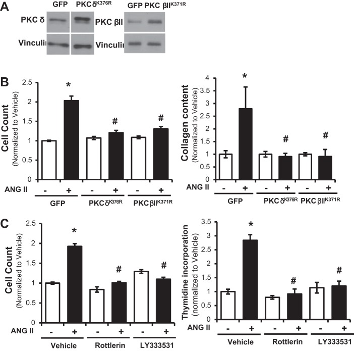
Pulmonary hypertension (PH) eventually leads to right ventricular (RV) fibrosis and dysfunction that is associated with increased morbidity and mortality. Although angiotensin II plays an important role in RV remodeling associated with hypoxic PH, the molecular mechanisms underlying RV fibrosis in PH largely remain unresolved. We hypothesized that PKC-p38 signaling is involved in RV collagen accumulation in PH and in response to angiotensin II stimulation. Adult male Sprague-Dawley rats were exposed to 3 wk of normoxia or hypoxia (10% FiO2 ) as a model of PH. Hypoxic rats developed RV hypertrophy and fibrosis associated with an increase in PKC βII and δ protein expression and p38 dephosphorylation in freshly isolated RV cardiac fibroblasts. Further mechanistic studies were performed in cultured primary cardiac fibroblasts stimulated with angiotensin II, a key activator of ventricular fibrosis in PH. Angiotensin II induced a reduction in p38 phosphorylation that was attenuated following chemical inhibition of PKC βII and δ. Molecular and chemical inhibition of PKC βII and δ abrogated angiotensin II-induced cardiac fibroblast proliferation and collagen deposition in vitro. The effects of PKC inhibition on proliferation and fibrosis were reversed by chemical inhibition of p38. Conversely, constitutive activation of p38 attenuated angiotensin II-induced increase of cardiac fibroblast proliferation and collagen accumulation. PKC βII- and δ-dependent inactivation of p38 regulates cardiac fibroblast proliferation and collagen deposition in response to angiotensin II, which suggests that the PKC-p38 signaling in cardiac fibroblasts may be involved and important in the pathophysiology of RV fibrosis in PH.

Keywords: PKC; angiotensin II; cardiac fibroblast; p38; pulmonary hypertension; right ventricle.


Figures

Fig. 1.
Fig. 1.
Pulmonary hypertension causes hypertrophy, collagen accumulation, and increased angiotensin II receptor expression in the right ventricle. A: weights of right and left ventricles (RV, LV) and interventricular septum (IVS) [normalized to body weight (BW)] in rats exposed to normoxic and hypoxic (10% FiO2) conditions (n = 6) for 3 wk. RV weight was also expressed as a ratio to LV plus IVS weight. B and C: Western blot analysis of equal protein (50 μg/lane) from tissue homogenates of RV, LV, and IVS from rats (n = 6) exposed to 3 wk of normoxia (N) or hypoxia (H) (10% FiO2) that were probed with an antibody specific to procollagen or angiotensin II receptor type 1 (AT1R). Data are expressed as ratio to vinculin and normalized to respective normoxic controls. *P < 0.05 vs. normoxia.
Fig. 2.
Fig. 2.
Differential PKC isoform expression and MAPK phosphorylation status in cardiac fibroblasts from rats with pulmonary hypertension. Western blot analysis of equal protein (50 μg/lane) from isolated RV (A, B, and E), LV (C), or IVS (D) cardiac fibroblast lysates of rats (n = 4) exposed to 3 wk of normoxia or hypoxia (10% FiO2) that were probed with antibodies specific to either PKC α, βII, δ, ϵ (A) or phosphorylated p38 or ERK1/2 and total p38 or ERK1/2 (BE). Data are calculated as a ratio of phosphorylated protein to total protein expressed and normalized to respective normoxic controls. *P < 0.05 vs. normoxia.
Fig. 3.
Fig. 3.
Angiotensin II-induced increase in cardiac fibroblast proliferation and collagen accumulation concomitant with enhanced PKC βII and δ expression and p38 dephosphorylation. Cardiac fibroblasts from healthy control rats that were treated with angiotensin II (1 μM) or vehicle for indicated times (0 to 2, 24, or 48 h), were assessed for p38 phosphorylation (n = 3) (A), PKCβII and δ expression (n = 4) (B), cell proliferation assessed by cell count (n = 6) and thymidine incorporation (n = 4) (C), and collagen content (n = 3) (D). Data are normalized to vehicle and presented as means ± SE; *P < 0.05 vs. vehicle, #P < 0.05 vs. 0-h time point.
Fig. 4.
Fig. 4.
Molecular and chemical inhibition of PKC βII and δ abolishes angiotensin II-induced increases in cardiac fibroblast proliferation and collagen accumulation. Cardiac fibroblasts from healthy control rats that were transfected with PKC βIIK371R and PKC δK376R, or green fluorescent protein (GFP) as control. Transfections were confirmed by Western blot analysis for PKC βII and δ (A). Transfected cardiac fibroblasts treated with angiotensin II (1 μM), or vehicle, for 48 h were measured for cell count (n = 7) (B) or collagen content (n = 5) (B). Cardiac fibroblasts preincubated with LY333531 (50 nM, 30 min) and rottlerin (3 μM, 30 min) followed by treatment with angiotensin II (1 μM), or vehicle, for 48 h were measured by cell count (n = 6) and thymidine incorporation (n = 4) (C). Data are normalized to vehicle and presented as means ± SE. *P < 0.05 vs. vehicle, #P < 0.05 vs. GFP, angiotensin II treatment.
Fig. 5.
Fig. 5.
Chemical inhibition of PKC βII and δ inhibits angiotensin II-induced dephosphorylation of p38. Cardiac fibroblasts from healthy control rats that were preincubated with LY333531 (50 nM, 30 min) and rottlerin (3 μM, 30 min), followed by treatment with angiotensin II (1 μM) or vehicle for 48 h, were assessed for p38 phosphorylation status and total p38 expression. Data are calculated as a ratio of phosphorylated protein to total protein expressed, or total protein to vinculin, and normalized to respective normoxic controls. Grouped data from 4 separate experiments are averaged and presented as means ± SE. *P < 0.05 vs. vehicle/vehicle, #P < 0.05 vs. vehicle/angiotensin II treatment.
Fig. 6.
Fig. 6.
Molecular activation of p38, via overexpression of p38 or the upstream activator MKK6, abolishes angiotensin II-induced increases in cardiac fibroblast proliferation and collagen accumulation. Cardiac fibroblasts from healthy control rats were transfected with wild-type p38 and MKK6 (p38wt and MKK6wt) or with GFP as control. Transfections were confirmed by Western blot analysis for p38 phosphorylation status and total p38 expression (A). Transfected cardiac fibroblasts treated with angiotensin II (1 μM), or vehicle, for 48 h were measured for cell count (n = 7) (B) and collagen content (n = 4) (C). Data are calculated as a ratio of phosphorylated protein to total protein expressed, or total protein to vinculin, and normalized to respective normoxic controls. Grouped data from 4 separate experiments are averaged and presented as means ± SE. *P < 0.05 vs. vehicle, #P < 0.05 vs. GFP, angiotensin II treatment.
Fig. 7.
Fig. 7.
Inhibition of p38 prevents the effect of PKC βII and δ inhibition on angiotensin II-induced proliferation and collagen content. Cardiac fibroblasts from healthy control rats that were preincubated with SB203580 (100 nM, 30 min), followed by treatment with LY333531 (50 nM, 30 min) and rottlerin (3 μM, 30 min), and angiotensin II (1 μM), or vehicle, for 48 h were measured for cell count (n = 9) (A) and collagen content (n = 5) (B). Data are normalized to vehicle and presented as means ± SE. *P < 0.05 vs. vehicle/vehicle, #P < 0.05 vs. vehicle/angiotensin II treatment.

References

    1. Bartha E, Solti I, Kereskai L, Lantos J, Plozer E, Magyar K, Szabados E, Kalai T, Hideg K, Halmosi R, Sumegi B, Toth K. PARP inhibition delays transition of hypertensive cardiopathy to heart failure in spontaneously hypertensive rats. Cardiovasc Res 83: 501–510, 2009. - PubMed
    1. Bogaard HJ, Abe K, Vonk Noordegraaf A, Voelkel NF. The right ventricle under pressure: cellular and molecular mechanisms of right-heart failure in pulmonary hypertension. Chest 135: 794–804, 2009. - PubMed
    1. Booz GW, Baker KM. Protein kinase C in angiotensin II signalling in neonatal rat cardiac fibroblasts. Role in the mitogenic response. Ann NY Acad Sci 752: 158–167, 1995. - PubMed
    1. Braun MU, LaRosee P, Simonis G, Borst MM, Strasser RH. Regulation of protein kinase C isozymes in volume overload cardiac hypertrophy. Mol Cell Biochem 262: 135–143, 2004. - PubMed
    1. Braz JC, Bueno OF, Liang Q, Wilkins BJ, Dai YS, Parsons S, Braunwart J, Glascock BJ, Klevitsky R, Kimball TF, Hewett TE, Molkentin JD. Targeted inhibition of p38 MAPK promotes hypertrophic cardiomyopathy through upregulation of calcineurin-NFAT signaling. J Clin Invest 111: 1475–1486, 2003. - PMC - PubMed

Publication types

MeSH terms

LinkOut - more resources