. 2015 Mar 27;427(6 Pt B):1375-1388.
doi: 10.1016/j.jmb.2015.01.019. Epub 2015 Feb 7.

The Pex1/Pex6 complex is a heterohexameric AAA+ motor with alternating and highly coordinated subunits

Affiliations

The Pex1/Pex6 complex is a heterohexameric AAA+ motor with alternating and highly coordinated subunits

Brooke M Gardner et al. J Mol Biol. .

Abstract

Pex1 and Pex6 are Type-2 AAA+ ATPases required for the de novo biogenesis of peroxisomes. Mutations in Pex1 and Pex6 account for the majority of the most severe forms of peroxisome biogenesis disorders in humans. Here, we show that the ATP-dependent complex of Pex1 and Pex6 from Saccharomyces cerevisiae is a heterohexamer with alternating subunits. Within the Pex1/Pex6 complex, only the D2 ATPase ring hydrolyzes ATP, while nucleotide binding in the D1 ring promotes complex assembly. ATP hydrolysis by Pex1 is highly coordinated with that of Pex6. Furthermore, Pex15, the membrane anchor required for Pex1/Pex6 recruitment to peroxisomes, inhibits the ATP-hydrolysis activity of Pex1/Pex6.

Keywords: AAA+ ATPase; Pex1; Pex15; Pex6; peroxisome.


Figures

Figure 1
Figure 1
A) Schematic representation of Pex1 and Pex6 from S. cerevisiae. Both are Type-2 AAA+ ATPases with an N-terminal domain (NTD) and two ATPase domains, D1 and D2. HR: N-terminal region homologous to the N-domain of p97 and NSF. A1 and A2: Walker A motif (GxxGxGKT) in the D1 or D2 ATPase domain. B1 and B2: Walker B motif (ϕϕϕϕDE, ϕ: hydrophobic amino acid) in the D1 or D2 ATPase domain. The alignment shows the conservation of the Walker A and Walker B motifs in ScPex1, ScPex6, ScCdc48, and ScSec18. Poorly conserved residues are underlined. B) Endogenous Pex1 and Pex6 from S. cerevisiae depend on the presence of nucleotide to form a complex. Pex6 co-immunoprecipitated with FLAG-tagged Pex1 in the presence of ATP or ATPγS. Pex1 co-immunoprecipitated with FLAG-tagged Pex6 in the presence of ATP, but this association is diminished when no nucleotide is present. A mock co-immunoprecipitation using the parent wild-type strain with untagged Pex1 and Pex6 served as a control. C) Recombinant Pex1-FLAG and His-Pex6 purified as a stoichiometric complex from E. coli. Pex1-FLAG and His-Pex6 were co-expressed in BL21* E. coli and subsequently purified by Ni-NTA agarose, anti-FLAG affinity resin, and size exclusion chromatography. The Superose 6 elution profile and SDS-PAGE analysis of the fractions show that the main peak contains both Pex1-FLAG and His-Pex6, and a minor peak represents a smaller homo-oligomer of Pex1-FLAG.
Figure 2
Figure 2
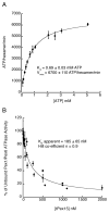
A) The recombinant Pex1-FLAG/His-Pex6 complex is an active ATPase with a Km of 0.7 mM ATP and Vmax of 6700 ATP/hexamer/min. B) The cytosolic domain of Pex15, tPex15, inhibits the ATPase activity of Pex1-FLAG/His-Pex6 with an apparent KD of 185 nM.
Figure 3
Figure 3
A) 2D class averages from negative-stain electron microscopy of recombinant Pex1-FLAG/His-Pex6 in the presence of 3 mM ATP. B) Three representative class averages of Pex1-FLAG/MBP-Pex6 show extra density for MBP at the apices of the heterohexamer, consistent with an attachment to the extended N-terminal domain. 2D classes were aligned and averaged to show the flexible attachment of the MBP tag to the N-terminal extensions of Pex6 in the heterohexamer. C) The 3D reconstruction of the Pex1-FLAG/His-Pex6 heterohexamer at 23-Å resolution. D1 and D2 mark the individual ATPase rings. The top view depicts the D1 ring and NTDs (left), and the bottom view shows the D2 ring (right). In the top view, the hash marks delineate the “dimer” interfaces in the “trimer of dimer” arrangement. D) The density of truncated NSF (EMDB: 2041) constituting only the D1 and D2 domains was docked into the 3D reconstruction of Pex1/Pex6 to emphasize the density of the Pex1/Pex6 N-terminal domains. E) The Pex1/Pex6 D2 ATPase ring exhibits the canonical architecture of the small α-helical AAA+ subdomain interacting with the large AAA+ subdomain of the counter-clockwise-neighboring subunit. Shown are homology models for the Pex1 and Pex6 D2 domains docked into the density of the Pex1/Pex6 D2 ATPase ring.
Figure 4
Figure 4
A) A proposed model showing the outlines for Pex1 and Pex6 in the segmented heterohexamer.
Figure 5
Figure 5
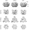
A comparison of the 3D reconstructions for ADP-, ATPγS-, and ATP-bound Pex1/Pex6 heterohexamer at 17, 23, and 23 Å resolution, respectively. A) Side-views of Pex1/Pex6 reveal no large changes in the N-terminal domain conformation. B) Vertical cross-sections at the axial pore show increased density in the D2-ring pore for the ATPγS-bound state. C) Horizontal cross-sections through the D1 ATPase domains. D) Horizontal cross-section through the D2 ATPase domains.
Figure 6
Figure 6
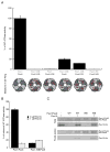
A) A comparison of ATPase activities for wild-type Pex1-FLAG/His-Pex6 and its variants with mutations in the D2 domains. The schematic below the graph indicates the D2 mutations made in the context of the heterohexamer, with Pex1 represented in light gray and Pex6 in dark gray. The ATP-binding sites are colored green when wild type and red when mutated. WB: Mutation of Glu to Asn in the Walker B motif, leading to inhibition of ATP hydrolysis. RK: Mutation of Arg to Lys in the R-finger, a residue contributed to the neighboring ATP-binding site, which inhibits ATP hydrolysis when mutated. B) The cytoplasmic domain of Pex15 inhibits the ATPase activity of the wild-type Pex1/Pex6 complex, but not the Pex1-WB/Pex6 mutant. C) The cytoplasmic domain of Pex15 co-immunoprecipitates with both wild-type and the D2 Walker B mutant Pex1/Pex6 complex. The immunoprecipitation was performed on purified proteins using the FLAG tag on Pex1.

Similar articles

Cited by

References

    1. Tabak HF, Braakman I, van der Zand A. Peroxisome formation and maintenance are dependent on the endoplasmic reticulum. Annu Rev Biochem. 2013;82:723–44. - PubMed
    1. Weller S, Gould SJ, Valle D. Peroxisome biogenesis disorders. Annu Rev Genomics Hum Genet. 2003;4:165–211. - PubMed
    1. Schluter A, Fourcade S, Ripp R, Mandel JL, Poch O, Pujol A. The evolutionary origin of peroxisomes: an ER-peroxisome connection. Mol Biol Evol. 2006;23:838–45. - PubMed
    1. Hoepfner D, Schildknegt D, Braakman I, Philippsen P, Tabak HF. Contribution of the endoplasmic reticulum to peroxisome formation. Cell. 2005;122:85–95. - PubMed
    1. Lam SK, Yoda N, Schekman R. A vesicle carrier that mediates peroxisome protein traffic from the endoplasmic reticulum. Proc Natl Acad Sci U S A. 2010;107:21523–8. - PMC - PubMed

Publication types

MeSH terms

Substances