. 2015 Mar 27;427(6 Pt B):1413-1427.
doi: 10.1016/j.jmb.2015.01.018. Epub 2015 Feb 7.

Elucidating the mechanism by which compensatory mutations rescue an HIV-1 matrix mutant defective for gag membrane targeting and envelope glycoprotein incorporation

Affiliations

Elucidating the mechanism by which compensatory mutations rescue an HIV-1 matrix mutant defective for gag membrane targeting and envelope glycoprotein incorporation

Philip R Tedbury et al. J Mol Biol. .

Abstract

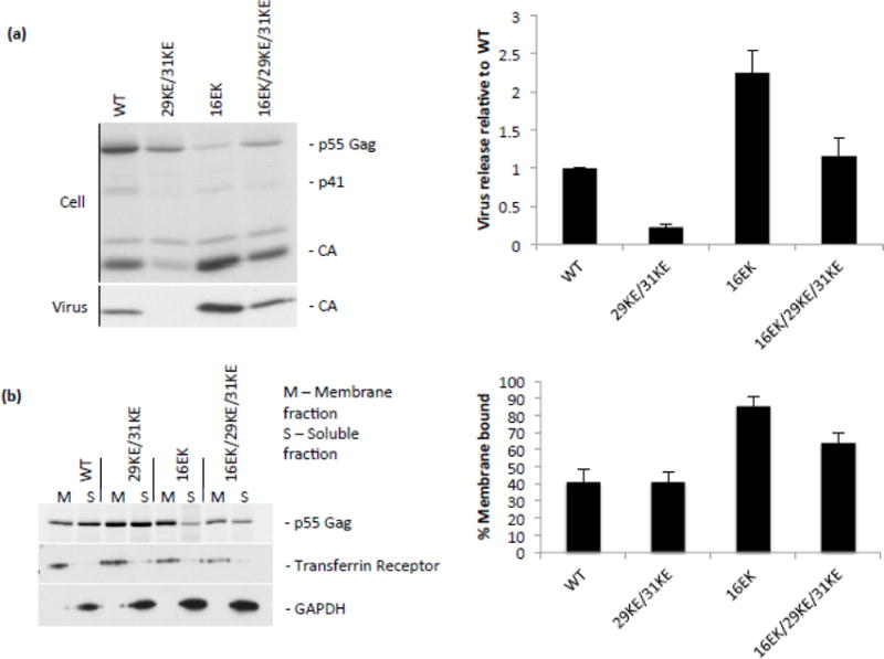
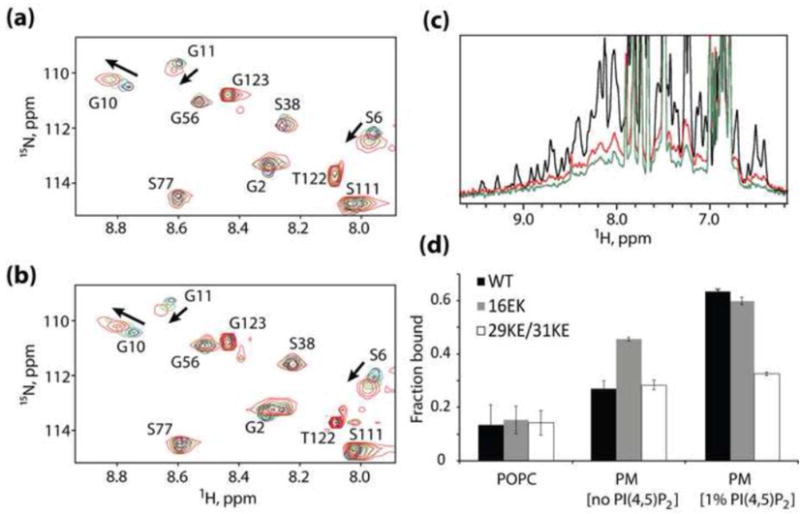
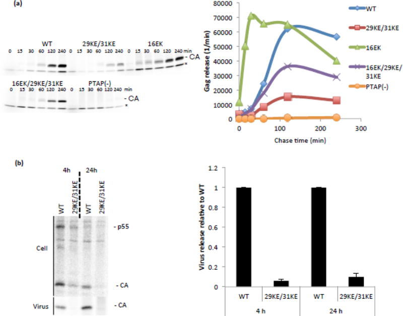
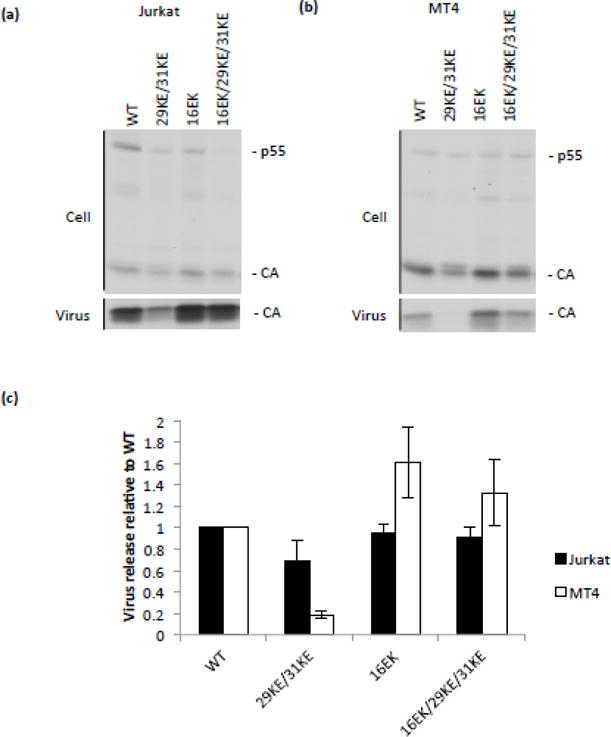
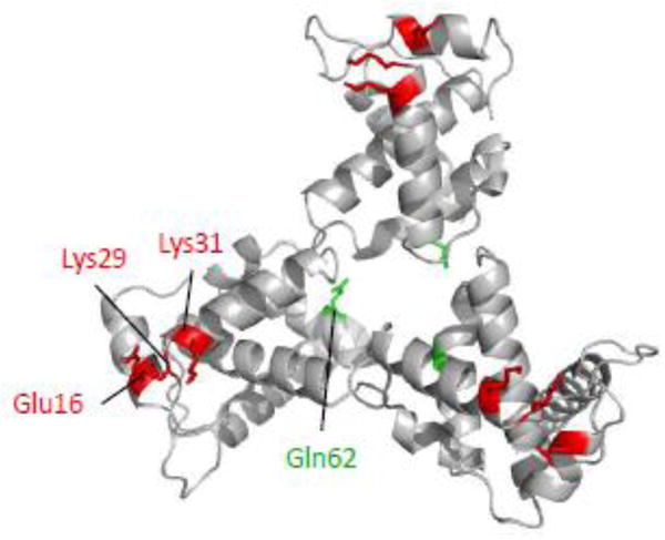
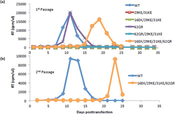
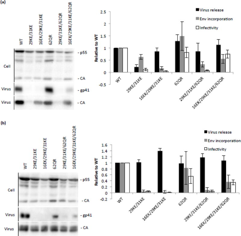
The matrix (MA) domain of the human immunodeficiency virus (HIV) 1 Gag is responsible for Gag targeting to the plasma membrane where virions assemble. MA also plays a role in the incorporation of the viral envelope (Env) glycoproteins and can influence particle infectivity post-maturation and post-entry. A highly basic region of MA targets Gag to the plasma membrane via specific binding to phosphatidylinositol-4,5-bisphosphate [PI(4,5)P2]. This binding also triggers exposure of an amino-terminal myristate moiety, which anchors Gag to the membrane. An MA mutant deficient for PI(4,5)P2 binding, 29KE/31KE, has been shown to mislocalize within the cell, leading to particle assembly in a multivesicular body compartment and defective release of cell-free particles in HeLa and 293T cells. Despite the defect in virus production in these cells, release of the 29KE/31KE mutant is not significantly reduced in primary T cells, macrophages and Jurkat T cells. 29KE/31KE virions also display an infectivity defect associated with impaired Env incorporation, irrespective of the producer cell line. Here we examine the properties of 29KE/31KE by analyzing compensatory mutations obtained by a viral adaptation strategy. The MA mutant 16EK restores virus release through enhanced membrane binding. 16EK also influences the infectivity defect, in combination with an additional MA mutant, 62QR. Additionally, the 29KE/31KE MA mutant displays a defect in proteolytic cleavage of the murine leukemia virus Env cytoplasmic tail in pseudotyped virions. Our findings elucidate the mechanism whereby an MA mutant defective in PI(4,5)P2 binding can be rescued and highlight the ability of MA to influence Env glycoprotein function.

Keywords: A-MLV; Gag; cytoplasmic tail; pseudotyping; retrovirus.


Figures

Figure 1
Figure 1
Virus release and membrane binding of 29KE/31KE and compensatory mutants. (a) HeLa cells were transfected with the HIV-1 mutants indicated, labelled with 35S Met/Cys for 3 h, then cell and virus lysates were immunoprecipitated and separated by SDS-PAGE. Samples from three independent experiments were analyzed by fluorography. Virion capsid, expressed as a percentage of total Gag, was plotted, +/− S.E.M.. (b) HeLa cells were transfected with the HIV-1 mutants indicated (in a PR virus), then labelled with 35S Met/Cys for 20 min. Cells were harvested by scraping, lysed by sonication and lysates placed at the bottom of a sucrose gradient. Following centrifugation, membrane-associated samples were collected from the top of the gradient, and soluble samples from the bottom. Gag was immunoprecipitated from these fractions and resolved by SDS-PAGE. Samples from four independent experiments were analyzed by fluorography. Gag percentage in the membrane-associated fraction was plotted +/− S.E.M. Flotation fractions were also analyzed by western blotting for transferrin receptor and GAPDH.
Figure 2
Figure 2
Mechanism of enhanced membrane binding by 16EK. (a,b) Concentration-dependent NMR chemical shift changes observed for residues S6, G10 and G11 in 1H-15N HSQC spectra of the wild-type (a) and 16EK (b) myrMA proteins. Spectral changes reflect a myristyl switch equilibrium switch from myristate-sequestered (low concentration) to myristate-exposed (high concentration) species [13], and indicate that the 16EK mutation does not perturb the myristyl switch. (c) One-dimensional 1H NMR spectra obtained for the myr-16EK mutant under various conditions: in the absence of liposomes (black) and in the presence of membrane mimetic liposomes that lack (red) or contain (green) PI(4,5)P2 (1%). Signal losses correlate with increasing fractions of liposome-bound protein. (d) Fractions of protein bound to POPC liposomes (left) and to membrane mimetic liposomes in the absence (center) and presence (right) of PI(4,5)P2 (1%), as determined by 1D 1H NMR. The wild type, 16EK, and 29KE/31KE proteins bind poorly to POPC liposomes but exhibit differentially enhanced binding to liposomes with PM-like compositions.
Figure 3
Figure 3
Virus release kinetics. (a) HeLa cells were transfected with the HIV-1 mutants indicated, labelled for 15 minutes with 35S Met/Cys then chased for 240 minutes. At the indicated times, media were replaced and virus harvested. Samples were separated by SDS-PAGE and analyzed by fluorography. For each time point the amount of Gag signal released per minute is plotted. Time courses were performed 4 times and data from a representative experiment is shown. A non-specific band is indicated with *. (b) HeLa cells were transfected with the HIV-1 mutants indicated, labelled with 35S Met/Cys, then cell and virus lysates were immunoprecipitated and separated by SDS-PAGE. Samples from three independent experiments were analyzed by fluorography. Virion capsid, expressed as a percentage of total Gag, was plotted relative to WT, +/− S.E.M.
Figure 4
Figure 4
Release of 29KE/31KE from T-cell lines. VSV-G pseudotyped viruses were generated by transfecting 293T cells with the HIV-1 mutants indicated and a VSV-G expression vector. These viruses were normalized by RT assay and used to infect Jurkat and MT4 T-cell lines. 24 h postinfection cells were labelled with 35S Met/Cys for 8 or 16 h, then cell and virus lysates were immunoprecipitated and separated by SDS-PAGE. (a) Jurkat. (b) MT4. (c) Samples from three independent experiments were analyzed by fluorography. Virion capsid, expressed as a percentage of total Gag, was plotted relative to WT, +/− S.E.M.
Figure 5
Figure 5
Model of the MA trimer showing residues Glu16, Lys29, Lys31 and Gln62. Model was generated in Pymol [49] based on Protein Data Bank coordinates 1HIW [25]. Gln62 (green) is found near the trimer interface, while the other residues (red) are located at the tips of the trimer.
Figure 6
Figure 6
Rescue of 29KE/31KE replication in Jurkats. (a) Jurkat T cells were transfected with the HIV-1 mutants indicated. Samples were taken every two days and the cells split 1:3. Samples were assayed for RT activity. (b) Virus supernatants from the peaks of WT and 16EK/29KE/31KE/62QR were collected, normalized by RT assay and used to reinfect Jurkat T cells for a 2nd passage. Replication assays were performed three times, data from a representative experiment are shown.
Figure 7
Figure 7
Virus release, Env incorporation and infectivity of 29KE/31KE and rescue mutations in HeLa and Jurkat cell lines. (a) HeLa cells were transfected with the HIV-1 mutants indicated. 48 h posttransfection virus and cell-associated samples were collected, separated by SDS-PAGE and analyzed by western blotting. Virus release was calculated as the amount of virion CA relative to total Gag levels, Env incorporation was expressed as amount of virion gp41 per virion CA. Virus-containing supernatants were used to infect TZM-bl cells; the resulting luciferase signal was normalized to the corresponding RT values to provide a measure of specific infectivity. Averages from four independent experiments are shown, +/− S.E.M. (b) 293T cells were transfected with the mutants indicated and a VSV-G expression vector. 48 h posttransfection supernatant was harvested and virus content determined by RT assay. These virus stocks were used to infect Jurkat T cells. 12 h postinfection the medium was replaced. After a further 48 h, virus- and cell-associated samples were harvested and analyzed as described in (a). Averages from three independent experiments are shown, +/− S.E.M.
Figure 8
Figure 8
Infectivity and pseudotyping 29/31KE and 16EK. (a) Viruses pseudotyped with VSV-G were generated by transfecting 293T cells with the HIV-1 mutants indicated and a VSV-G expression vector. These viruses were normalized by RT assay and used to infect Jurkat and MT4 T-cell lines. 48 h post-infection virus was collected and RT assays and TZM-bl infections performed. Virus-containing supernatants were used to infect TZM-bl cells; the resulting luciferase signal was normalized to the corresponding RT values to provide a measure of specific infectivity. Means of three independent experiments are plotted, +/− S.E.M. (b) HeLa cells were transfected with Env-deficient clones of the HIV-1 mutants and viral glycoproteins indicated. After 24 h, virus infectivity was determined as in (a). (c) HeLa cells were transfected with Env-deficient clones of the HIV-1 mutants indicated and A-MLV Env. After 48 h, virus was pelleted by ultracentrifugation and analyzed by western blotting. (d) HeLa cells were transfected with Env-deficient clones of the HIV-1 mutants indicated and AMLV p12* Env. After 24 h, virus infectivity was determined as in (a). Means of three independent experiments are plotted +/− S.E.M.

Similar articles

Cited by

References

    1. Sundquist WI, Kräusslich H-G. HIV-1 Assembly, Budding, and Maturation. Cold Spring Harb Perspect Med. 2012;2:a006924. doi: 10.1101/cshperspect.a006924. - DOI - PMC - PubMed
    1. Ganser BK, Li S, Klishko VY, Finch JT, Sundquist WI. Assembly and analysis of conical models for the HIV-1 core. Science. 1999;283:80–3. - PubMed
    1. Lu K, Heng X, Summers MF. Structural determinants and mechanism of HIV-1 genome packaging. J Mol Biol. 2011;410:609–33. doi: 10.1016/j.jmb.2011.04.029. - DOI - PMC - PubMed
    1. Bell NM, Lever AML. HIV Gag polyprotein: processing and early viral particle assembly. Trends Microbiol. 2013;21:136–44. doi: 10.1016/j.tim.2012.11.006. - DOI - PubMed
    1. Votteler J, Sundquist WI. Virus budding and the ESCRT pathway. Cell Host Microbe. 2013;14:232–41. doi: 10.1016/j.chom.2013.08.012. - DOI - PMC - PubMed

Publication types

MeSH terms

Substances