. 2015 Jun;172(12):3072-85.
doi: 10.1111/bph.13111. Epub 2015 Apr 10.

Pharmacological modulation of autophagy to protect cardiomyocytes according to the time windows of ischaemia/reperfusion

Affiliations

Pharmacological modulation of autophagy to protect cardiomyocytes according to the time windows of ischaemia/reperfusion

Qiulin Xu et al. Br J Pharmacol. 2015 Jun.

Abstract

Background and purpose: Targeted modulation of autophagy induced by myocardial ischaemia/reperfusion has been the subject of intensive investigation, but it is debatable whether autophagy is beneficial or harmful. Hence, we evaluated the effects of pharmacological manipulation of autophagy on the survival of cardiomyocytes in different time windows of ischaemia/reperfusion.

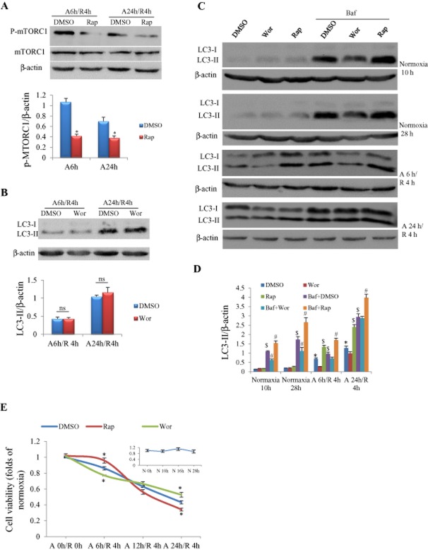
Experimental approach: We examined the autophagy and apoptosis in cardiomyocytes subjected to different durations of anoxia/re-oxygenation or ischaemia/reperfusion, and evaluated the effects of the autophagic enhancer rapamycin and inhibitor wortmannin on cell survival.

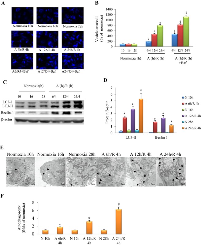
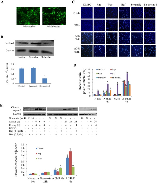
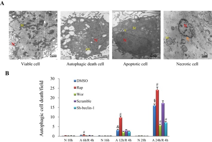
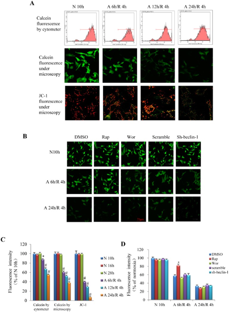
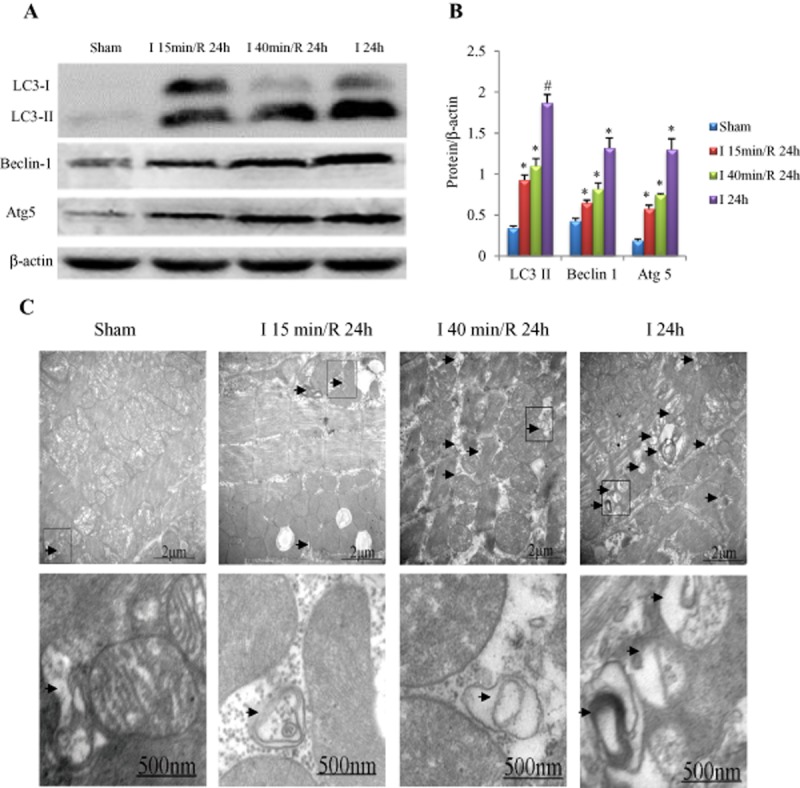
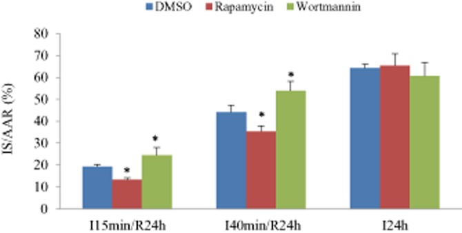
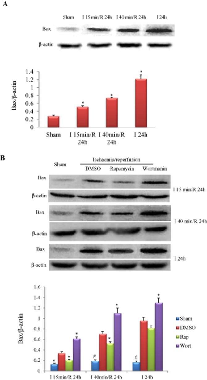
Key results: In neonatal rat cardiomyocytes (NRCs) or murine hearts, autophagy was increased in response to anoxia/reoxygenation or ischaemia/reperfusion in a time-dependent manner. Rapamycin-enhanced autophagy in NRCs led to higher cell viability and less apoptosis when anoxia was sustained for ≦ 6 h. When anoxia was prolonged to 12 h, rapamycin did not increase cell viability, induced less apoptosis and more autophagic cell death. When anoxia was prolonged to 24 h, rapamycin increased autophagic cell death, while wortmannin reduced autophagic cell death and apoptosis. Similar results were obtained in mice subjected to ischaemia/reperfusion. Rapamycin inhibited the opening of mitochondrial transition pore in NRCs exposed to 6 h anoxia/4 h re-oxygenation but did not exert any effect when anoxia was extended to 24 h. Similarly, rapamycin reduced the myocardial expression of Bax in mice subjected to short-time ischaemia, but this effect disappeared when ischaemia was extended to 24 h.

Conclusions and implications: The cardioprotection of autophagy is context-dependent and therapies involving the modification of autophagy should be determined according to the duration of ischaemia/reperfusion.


Figures

Figure 1
Figure 1
Autophagy triggered by A/R in cultured NRCs. (A) Autophagic activity detected by MDC stain in the presence/absence of bafilomycin A1 (Baf, 0.1 μM). (B) Area of MDC-positive vesicle per cell, n = 6 in each group. (C) Examples of Western blotting picture of autophagy-related proteins. (D) Quantitative analysis of LC3-II, and beclin-1; n = 5, in each group. (E) Autophagic vesicle (arrow) increased in an anoxia duration-dependent manner. (F) Quantitative analysis of autophagic vacuole (the number of autophagosome per optical field, 30 optical fields were used to calculate and results standardized to the responding normoxia group), n = 5 in each group. In (B, D and F), *P < 0.05 versus the corresponding normoxia group; #P < 0.05 versus A 6 h/R 4 h group; §P < 0.05 versus the corresponding A/R group without bafilomycin A1 treatment. N, normoxia.
Figure 2
Figure 2
Influences of autophagy manipulation on autophagic flux and viability of NRCs in response to A/R. (A) Rapamycin (Rap) at 0.1 μM significantly inhibited phosphorylation of mTOR in cardiomyocytes with A/R insults. *P < 0.05, n = 5. (B) Wortmannin (Wor) at 3 nM had no effect on LC3-II protein expression. Experiments were repeated five times; ns, not significant. (C) Representative pictures of Western blot for LC3-II in response to A/R with/without autophagy manipulation. (D) Semi-quantification of LC3-II protein expression as shown in (C). Rapamycin = 0.1 μM; wortmannin = 0.2 μM; bafilomycin A1 (Baf) = 0.1 μM. *P < 0.05 versus the DMSO group at the corresponding time point of normoxia; #P < 0.05 versus the bafilomycin A1 + DMSO group at the same time point; $P < 0.05 versus the DMSO group at the same time point, n = 5. (E) Viability of cardiomyocytes measured by MTT assay. *P < 0.05, n = 24, 6 and 6 in control (DMSO), rapamycin and wortmannin groups respectively. The insert figure shows cell viability in response to different normoxia duration, n = 6 at each time point.
Figure 3
Figure 3
Effects of pharmacological and genetic manipulation of autophagy on apoptosis in cultured NRCs with different durations of A/R. (A) Infective efficiency of adenovirus carrying shRNA for beclin-1 (Ad-sh-beclin-1) or control (Ad-scramble) in cultured cardiomyocytes for 48 h detected by the green fluorescence of co-expressed EGFP, multiple of infection = 5. (B) Western blot analysis of beclin-1 protein levels in response to Ad-sh-beclin-1 or Ad-scramble infection (*P < 0.01 vs. scramble group. The experiment was repeated five times). (C) Representative Hoechst staining pictures under microscope. The bright blue-stained nucleolus represent apoptotic cells. (D) Apoptotic rate calculated from five fields of view for each group. &P < 0.05 versus the corresponding DMSO group under normoxia (N 10 h or N 28 h), *P < 0.05 versus the corresponding DMSO group. #P < 0.05 versus the corresponding scramble group. Experiments were repeated five times. (E) Western blotting results of cleaved caspase 3. #P < 0.05 versus the corresponding DMSO group under normoxia (N 10 h or N 28 h), *P < 0.05 versus the corresponding DMSO group. Experiments were repeated five times. Concentration used: wortmannin (Wor) 0.2 μM; rapamycin (Rap) 0.1 μM; bafilomycin A1 (Baf) 0.1 μM. N, normoxia; Re-oxy, reoxygenation.
Figure 4
Figure 4
Influence of A/R and autophagic manipulation on autophagic cell death in NRCs. (A) Examples of viable, autophagic, apoptotic and necrotic cardiomyocytes under electronic microscopy. N, nucleus; M, mitochondria; AV, autophagic vacuole; arrow, disruption of plasma membrane. (B) Number of cells detected undergoing autophagic death in response to different severities of A/R and treatment with rapamycin (Rap, 0.1 μM) or wortmannin (Wor, 0.2 μM) or adenovirus carrying sh-beclin 1 or scramble. &P < 0.05 versus the DMSO group with insult of A 6 h/R 4 h; §P < 0.05 versus the DMSO group with insult of A 12 h/R 4 h; #P < 0.05 versus the DMSO group with the same insult, n = 5 in each group. N, normoxia.
Figure 5
Figure 5
Influence of different A/R injury on the opening of mPTP. (A) Representative pictures of calcein fluorescence detected with flow cytometry (first line) and laser confocal microscopy (second line) as well as JC-1 fluorescence (reflecting the depolarization of mitochondria) detected by laser confocal microscopy (third line). (B) Representative pictures of calcein fluorescence in response to pharmacological or genetic manipulation of autophagy. (C) Quantitative analysis of fluorescent intensity in the mitochondria of cardiomyocytes as shown in A. *P < 0.05, #P < 0.01 (anova) versus normoxia group. (D) Quantitative analysis of calcein fluorescent intensity in the mitochondria of cardiomyocytes as shown in B. *P < 0.05 versus DMSO group with the same A/R insult. n = 6 in each group of calcein flow cytometer assay, calcein confocal and JC-1 analysis respectively. Rap, rapamycin; Wor, wortmannin; N, normoxia.
Figure 6
Figure 6
Influence of ischaemia duration on myocardial autophagy. (A) Examples of Western blotting picture of autophagy-related proteins. (B) Quantitative analysis of LC3-II, beclin-1 and Atg5. *P < 0.05, #P < 0.01 versus sham, n = 5 in each group. (C) Electronic microscope findings. Autophagic vesicles (arrow) increased in an ischaemia duration-dependent manner (the magnified autophagic vesicles are shown in the second line).
Figure 7
Figure 7
Influence of ischaemia duration on myocardial injury. Myocardial infarct size (IS) was reduced by rapamycin (0.25 mg·kg−1) and increased by wortmannin (0.6 mg·kg−1) when ischaemia duration persisted for 15 or 40 min, but this effect disappeared when the ischaemia duration was extended to 24 h. For both (A and B), *P < 0.05 versus the responding control group (DMSO), n = 8 in each group.
Figure 8
Figure 8
Influence of ischaemia duration and autophagy manipulation on Bax expression. (A) Bax was increased in an ischaemia duration-dependent manner. *P < 0.05 versus sham, n = 5 in each group. (B) Effects of rapamycin (0.25 mg·kg−1) or wortmannin (0.6 mg·kg−1) on myocardial Bax expression in mice subjected to different durations of I/R injury. *P < 0.05, #P < 0.01 versus the corresponding DMSO-treated group, n = 5 in each group.

References

    1. Aki T, Yamaguchi K, Fujimiya T, Mizukami Y. Phosphoinositide 3-kinase accelerates autophagic cell death during glucose deprivation in the rat cardiomyocyte-derived cell line H9c2. Oncogene. 2003;22:8529–8535. - PubMed
    1. Alexander SPH, Benson HE, Faccenda E, Pawson AJ, Sharman JL, Spedding M, et al. The Concise Guide to PHARMACOLOGY 2013/14: Enzymes. Br J Pharmacol. 2013;170:1797–1867. - PMC - PubMed
    1. Baines CP, Kaiser RA, Sheiko T, Craigen WJ, Molkentin JD. Voltage-dependent anion channels are dispensable for mitochondrial-dependent cell death. Nat Cell Biol. 2007;9:550–555. - PMC - PubMed
    1. Cao X, Chen A, Yang P, Song X, Liu Y, Li Z, et al. Alpha-lipoic acid protects cardiomyocytes against hypoxia/reoxygenation injury by inhibiting autophagy. Biochem Biophys Res Commun. 2013;441:935–940. - PubMed
    1. Carreira RS, Lee Y, Ghochani M, Gustafsson AB, Gottlieb RA. Cyclophilin D is required for mitochondrial removal by autophagy in cardiac cells. Autophagy. 2010;6:462–472. - PMC - PubMed

Publication types

MeSH terms