. 2015 Feb 18;85(4):726-41.
doi: 10.1016/j.neuron.2015.01.008. Epub 2015 Feb 5.

N17 Modifies mutant Huntingtin nuclear pathogenesis and severity of disease in HD BAC transgenic mice

Affiliations

N17 Modifies mutant Huntingtin nuclear pathogenesis and severity of disease in HD BAC transgenic mice

Xiaofeng Gu et al. Neuron. .

Abstract

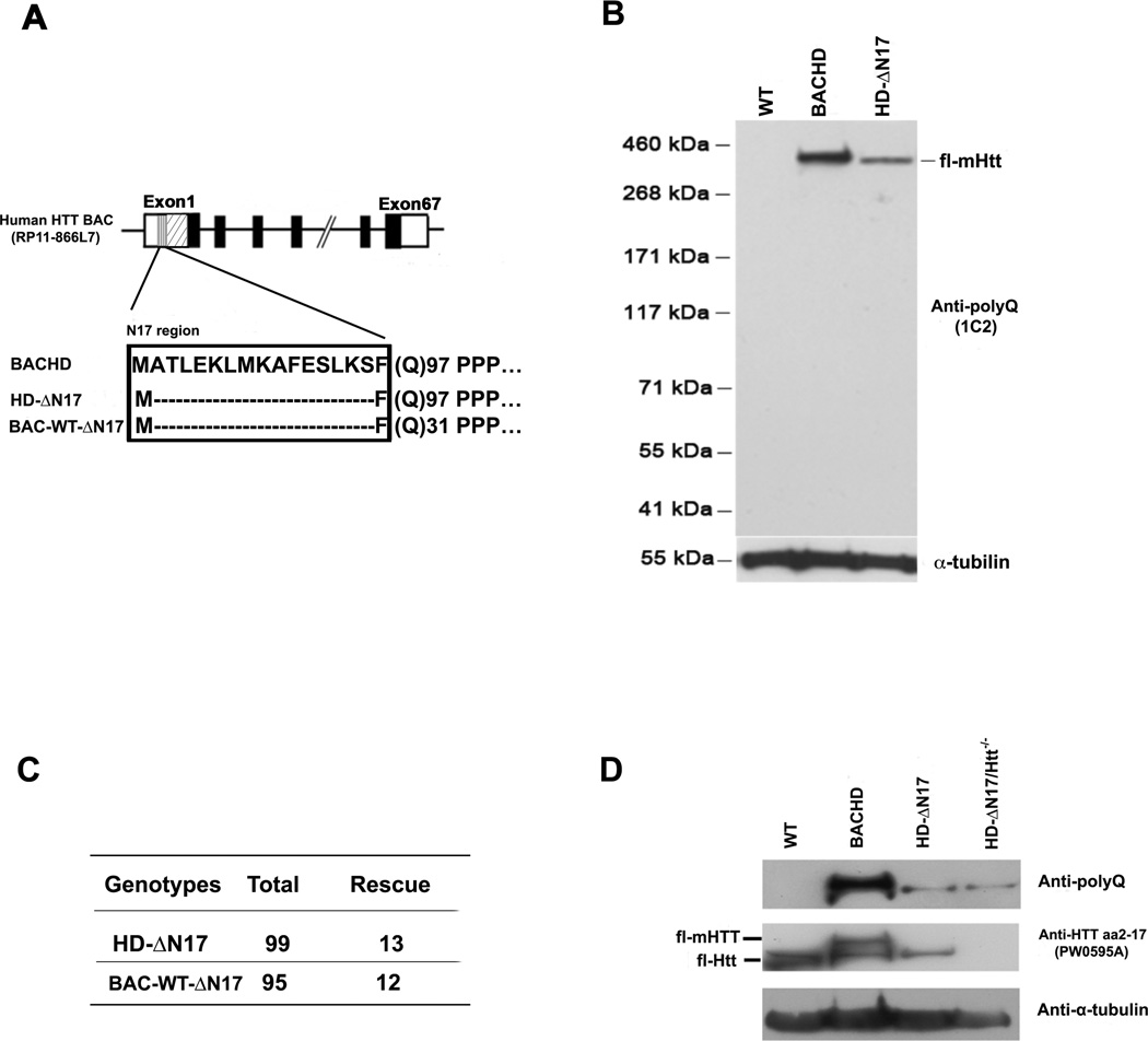
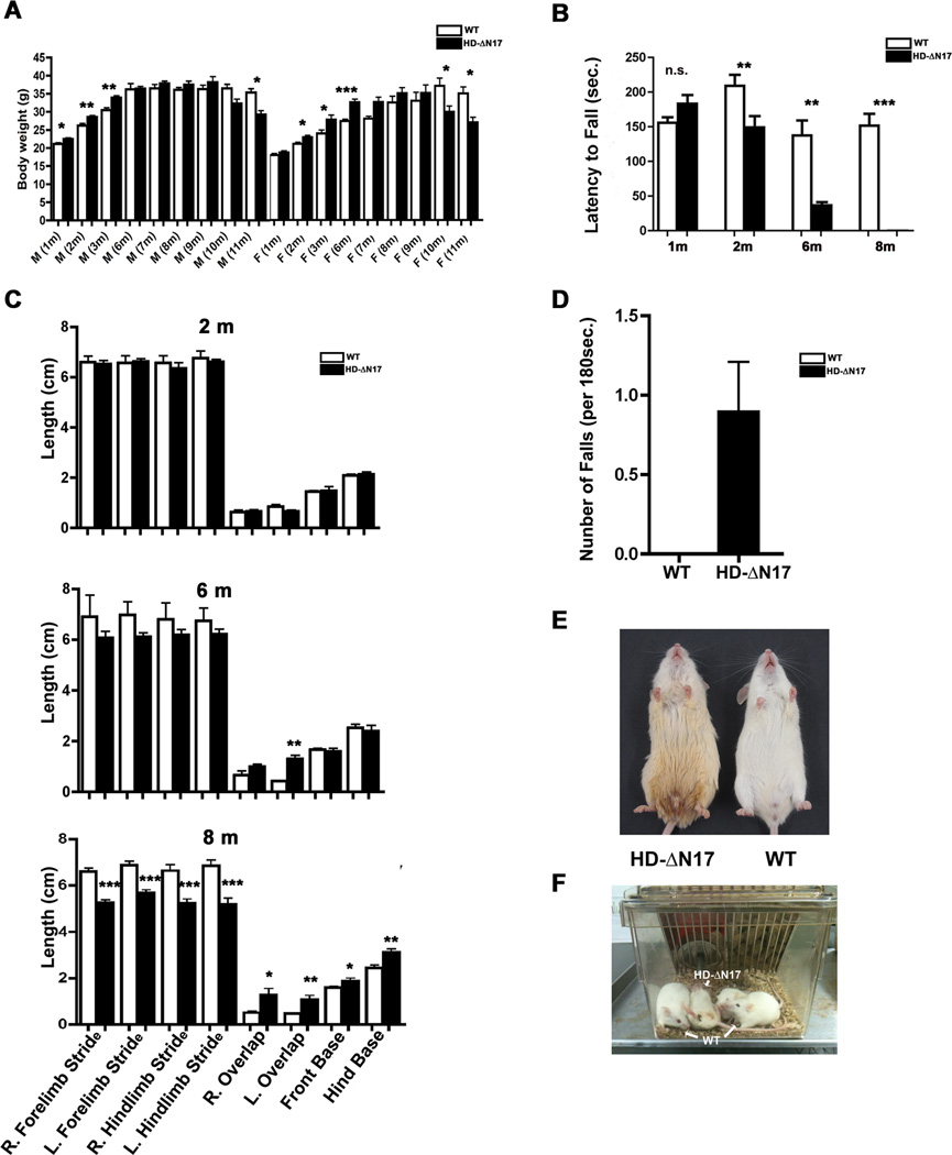
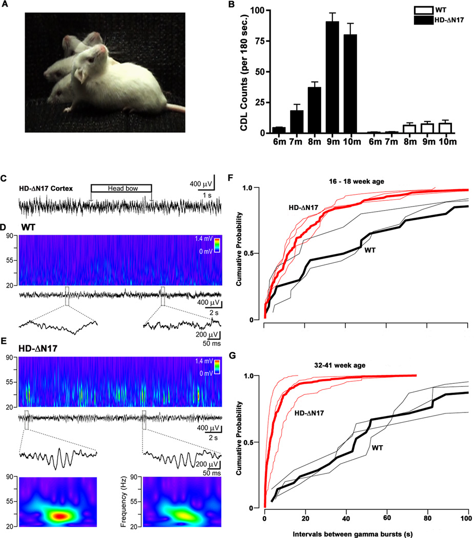
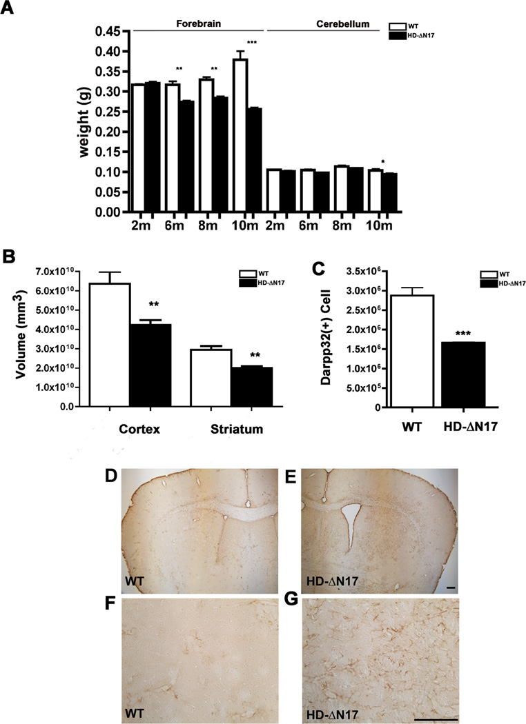
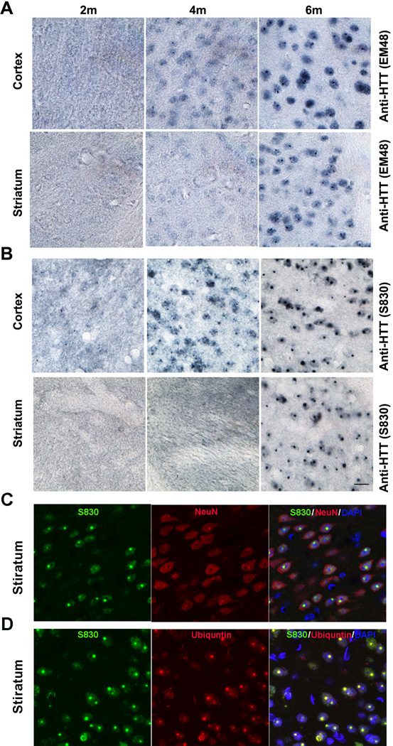
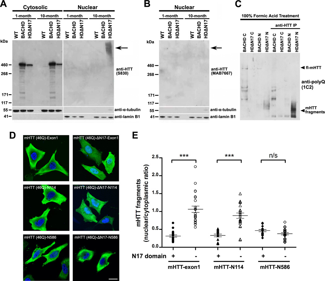
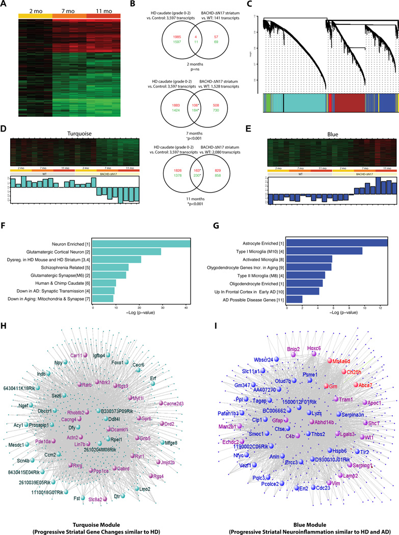
The nucleus is a critical subcellular compartment for the pathogenesis of polyglutamine disorders, including Huntington's disease (HD). Recent studies suggest the first 17-amino-acid domain (N17) of mutant huntingtin (mHTT) mediates its nuclear exclusion in cultured cells. Here, we test whether N17 could be a molecular determinant of nuclear mHTT pathogenesis in vivo. BAC transgenic mice expressing mHTT lacking the N17 domain (BACHD-ΔN17) show dramatically accelerated mHTT pathology exclusively in the nucleus, which is associated with HD-like transcriptionopathy. Interestingly, BACHD-ΔN17 mice manifest more overt disease-like phenotypes than the original BACHD mice, including body weight loss, movement deficits, robust striatal neuron loss, and neuroinflammation. Mechanistically, N17 is necessary for nuclear exclusion of small mHTT fragments that are part of nuclear pathology in HD. Together, our study suggests that N17 modifies nuclear pathogenesis and disease severity in HD mice by regulating subcellular localization of known nuclear pathogenic mHTT species.


Figures

Figure 1
Figure 1. Generation and Characterization of BAC Mice Expressing ΔN17 Forms of Huntingtin
(A) Schematic of transgene construction depicting the constructs used in generating BACHD-ΔN17 and BAC-WT-ΔN17 mice. (B) Brain lysates of WT, BACHD and BACHD-ΔN17 mice were probed with anti-polyQ antibody to determine mHTT expression levels. The anti-α-tubulin antibody was used for loading control. (1C2). (C) BACHD-ΔN17 and BAC-WT-AN17 transgene can rescue embryonic lethality of homozygous Htt null mice. The panel shows the number of rescue mice among the total number of mice born. (D) An anti-HTT aa2-17 (PW0595A) antibody detects the N17 domain both human HTT and murine Htt. The anti-polyQ (1C2) antibody detects only human mHTT. Brain lysates of WT, BACHD, BACHD-ΔN17 and BACHD-ΔN17/Htt−/− mice were probed with 1C2 and anti-N17 (PW0595A) antibodies, with anti-α-tubulin as loading control. See also Figures S1.
Figure 2
Figure 2. Age-Dependent Motor Deficits and General Health Decline in BACHD-ΔN17 Mice
(A) Body weights of both male (M) and female (F) BACHD-ΔN17 mice and WT mice from 1m to 11m of age. Significant interaction between genotypes and age (F=3.32, p<0.01) for females and age (F=31.62, p<0.001) for males was detected (two-way ANOVA). Further Student’s t-tests at each age showed that both females and males transgenic mice gained weight before 6 months of age, before losing significant weight around 10 months of age (*p<0.05, **p<0.01, ***p<0.001). (B) Motor performance of BACHD-ΔN17 mice and WT littermates was measured using an accelerating rotarod test. (C) Gait analyses were performed at 2m, 6m and 8m of age (*p<0.05, **p<0.01, Student’s t-tests). (D) Spontaneous falls were counted in BACHD-ΔN17 mice and WT littermates at 10m of age over a period of 180 seconds. (E and F) BACHD-ΔN17 mice show unkempt fur and urine scalding at 10m of age (E) and lay upside-down in the home cage (F). Data are shown as mean ± SEM. Student’s t-tests were performed to compare the results of BACHD-ΔN17 and WT mice (*p<0.05; **p<0.01; ***p<0.001). See also Figures S2, S6, S7.
Figure 3
Figure 3. Progressive Chorea/Dystonia-Like Movement Deficits and Aberrant Striatal Neuronal Activities in BACHD-ΔN17 Mice
(A) Composited image depicting chorea/dystonia-like (CDL) head movement in BACHD-ΔN17 mouse. (B) Quantification of CDLs in 3 minutes of video recording. Two-way ANOVA reveals significant difference between BACHD-ΔN17 and WT littermates in age and genotype interaction: F=16.55, p<0.0001; phenotype: F=117.2, p<0.0001. Data are shown as mean ± SEM. (C) Cortical field potential recording showing no epileptic activity during chorea-like movement (horizontal box over the recording) in an 8-month-old BACHD-ΔN17 mouse. (D) There is no significant gamma activity in the dorsal striatum of WT littermates. Middle: local field potential recording in the dorsal striatum of a WT littermate. Top: corresponding wavelet transform shows minor activity in the low gamma band (20–50 Hz). Bottom: enlarged segments of the field potential recordings indicated by dashed boxes. (E) Frequent gamma events in BACHD-ΔN17 mice. From top second row: local field potential recording in the dorsal striatum. Top: corresponding wavelet transform shows transient increments in the gamma range. Third row: sample large gamma events, enlarged segments of the local field potential recording indicated by dashed boxes with the corresponding wavelet transform (bottom). (F and G) Progression of the large gamma events in the striatum with aging in BACHD-ΔN17 mice. Cumulative probability plot of inter-event interval in younger (F) and older (G) animals. Red: transgenic, black: wild type, thicker lines indicate the corresponding pooled cumulative probability. See also Video clips 1 to 5, Figures S6, S7.
Figure 4
Figure 4. Progressive and Selective Brain Atrophy, Striatal Neuronal Cell Loss, and Gliosis in BACHD-ΔN17 Mice
(A) Weight of the forebrain and cerebellum of BACHD-ΔN17 and WT littermates was measured at 2, 6, 8 and 10 months of age. Progressive forebrain weight loss is detected in BACHD-ΔN17 mice starting at 6 months of age and progresses between 6m and 10m of age Two-way ANOVA reveals significant differences between HD-AN17 and WT littermates in age and genotype interaction (F=49.94, p<0.0001; genotype: F=173.2, p<0.0001). No weight-loss is detected in the cerebellum until 10m of age. (B) Unbiased stereological measurement of brain regions was performed to measure the cortical and striatal volume in BACHD-ΔN17 mice and WT littermates. (C). Unbiased stereological counting revealed robust loss of Darpp32+ neurons in 10m old BACHD-ΔN17 mice compared to WT controls. (D – G) Reactive astrogliosis is detected in striatum and deep cortical layers of BACHD-ΔN17 mouse brain with GFAP immunohistochemical staining (E, G). No such gliosis is seen in control mouse brain (D, F). Data are shown as mean ± SEM. Student’s t-tests were performed to compare BACHD-ΔN17 and WT samples (*p<0.05; **p<0.01; ***p<0.001). Scale bar = 100 µm. See also Figures S3, S6, S7.
Figure 5
Figure 5. Deletion of N17 Selectively Accelerates mHTT Nuclear Accumulation and Aggregation in BACHD-ΔN17 Mouse Brains
(A and B) Immunohistochemistry with EM48 (A) and S830 (B) antibodies to detect mHTT nuclear accumulation and aggregation in 2m, 4m and 6M old BACHD-ΔN17 mouse cortex and striatum. (C). Immunofluorescent staining to simultaneously detect S830+ mHTT aggregates (green), NeuN+ neurons (red), and merged signals with DAPI nuclear staining (Yellow and Purple). (D). Immunofluorescent staining to simultaneously detect S830+ mHTT aggregates (green), Ubiquitin+ nuclear inclusions (red), and merged signals with DAPI nuclear staining (Yellow and Purple). Scale bar = 50 µm. See also Figures S4, S6, S7.
Figure 6
Figure 6. N17-Dependent Nuclear Accumulation of mHTT In Vivo and In Vitro
(A and B) Western blot analysis of cytosolic and nuclear fractions from the forebrain extracts from 1m and 10m old WT, BACHD, and BACHD-ΔN17 mice. The blot was probed with antibodies against (A) the N-terminal human mHTT exon-1 (S830), and (B) the C-terminal HTT (MAB7667; Shirasaki et al., 2012). Lamin B1 and α-actin served as loading controls for nuclear and cytosolic fractions, respectively. (C) Nuclear (N) and cytosolic (C) extracts of forebrains from 1 and 10 month old WT, BACHD, and BACHD-ΔN17 were treated with 100% formic acid to dissolve the SDS-insoluble mHTT aggregates (Lunkes et al, 2002), followed by Western analysis. For better detecting the presence of mHTT fragments shown in lanes 5–8, mHTT were immunoprecipitated and detected by an anti-polyQ antibody (1C2). (D) HEK-293 cells were transfected with plasmids carrying mHTT fragments of different sizes, i.e. Exon1, CpA (N114), Caspase 6 (N586), and either with (left) or without N17 (right, deletion of amino acid residues 2–16). All constructs of fragments were fused at C-terminal with HA-tag and are as labeled on the images. The subcellular localization of mHTT was revealed by immunostaining of the HA-tag and imaged by confocal microscopy. (E) The nuclear and cytosolic distribution of mHTT fragments in (D) was quantified based on an established protocol (Ch'ng et al., 2012). The ratios of mean intensity of nuclear and cytosolic fluorescent signals of individual cells were plotted and analyzed using Student’s t test. *** p < 0.001; Scale bar = 20 µm. See also Figure S8.
Figure 7
Figure 7. Progressive HD-like Transcriptional Dysregulation and WGCNA Analyses of Gene Expression Profiles in BACHD-ΔN17 Striatum
(A) Heat map representing differentially expressed genes at different ages in the striatum of BACHD-ΔN17 compared to WT mice. Up-regulated genes are shown in red and down-regulated genes in green; color intensity corresponds to fold change in expression. (B) Venn diagrams representing the overlap between genes previously reported as dysregulated in HD patients (Kuhn et al., 2007, left circles) and genes dysregulated in the BACHD-ΔN17 model (this study, right circles). An increasing degree of overlap between the two datasets is observed in both up-regulated (red numbers) and down-regulated (green numbers) genes as the BACHD-ΔN17 animals age (p = hypergeometric test). (C) Cluster dendrogram generated by hierarchical clustering of probes on the basis of topological overlap. Seven modules of highly co-expressed transcripts were identified using a dynamic cutting algorithm, and color-coded modules (Blue, Red, Green, Turquoise, Brown, and Pink respectively). (D) Heatmap and first principal component of expression data for genes in the Turquoise and Blue modules identified age and genotype-dependent, coordinated changes in gene expression within these two modules. (E) Enrichment analyses indicate overrepresentation of neuronal genes and genes implicated in other neurodegenerative disorders (e.g. schizophrenia, AD) in the Turquoise module, and of gene transcripts related to neuroinflammation, including microglia and astrocyte markers, in the Blue module. (F) Network visualization of the Turquoise and Blue modules with top hubs highlighted (inner circle). Genes already shown to have transcriptional changes in HD caudate (Hodges et al., 2006) are depicted in purple. Genes involved in AD and frontal temporal dementia (e.g. Grn) are depicted in red.
Figure 8
Figure 8. A Working Model of N17 Domain Role in Regulating Nuclear Pathogenesis in HD
In HD neurons, full-length or fragmented mHTT species are predominantly located in the cytoplasm due to N17 function in Crm1-dependent nuclear export and cytoplasmic membrane association. For nuclear pathogenesis to occur, two events are likely needed to occur. First, the generation of small mHTT N-terminal fragments (e.g. mHTT-exon1 or CpA), which are known to be part of nuclear mHTT pathology in HD brains. Since these small mHTT fragments have functional N17, they are located predominantly in the cytoplasm, where N17 can facilitate their clearance (Thompson et al., 2009). The various mHTT species can also elicit cytoplasmic pathogenesis. For nuclear pathogenesis to occur, N17 domain on the small mHTT fragments has to be impaired either functionally (e.g. oligomerization or aggregation of mHTT fragments in the nucleus) or physically (e.g. deleterious N17 PTMs or truncation/proteolysis of N17 itself). Future studies are needed to determine whether N17 impairment can be demonstrated among the nuclear accumulated mHTT species in HD patients or mouse models. Moreover, the molecular pathways that may regulate N17 function in nucleocytoplasmic trafficking and whether manipulating these pathways could modify nuclear mHTT pathogenesis and overall disease phenotypes in HD models or HD remain to be elucidated.

References

    1. Atwal RS, Desmond CR, Caron N, Maiuri T, Xia J, Sipione S, Truant R. Kinase inhibitors modulate huntingtin cell localization and toxicity. Nature Chemical Biology. 2011;7:453–452. - PubMed
    1. Atwal RS, Xia J, Pinchev D, Taylor J, Epand RM, Truant R. Huntingtin has a membrane association signal that can modulate huntingtin aggregation, nuclear entry and toxicity. Human Molecular Genetics. 2007;16:2600–2615. - PubMed
    1. Benn CL, Landles C, Li H, Strand AD, Woodman B, Sathasivam K, Li SH, Ghazi-Noori S, Hockly E, Faruque SM, et al. Contribution of nuclear and extranuclear polyQ to neurological phenotypes in mouse models of Huntington's disease. Hum Mol Genet. 2005;14:3065–3078. - PubMed
    1. Björkqvist M, Wild EJ, Thiele J, Silvestroni A, Andre R, Lahiri N, Raibon E, Lee RV, Benn CL, Soulet D, et al. A novel pathogenic pathway of immune activation detectable before clinical onset in Huntington's disease. J Exp Med. 2008;205:1869–1877. - PMC - PubMed
    1. Burgess DL, Noebels JL. Single gene defects in mice: the role of voltage-dependent calcium channels in absence models. Epilepsy Res. 1999;36:111–122. - PubMed

Publication types

MeSH terms

Associated data