

Morphological and positional changes of the carpal arch and median nerve during wrist compression
- PMID: 25661267
- PMCID: PMC4363297
- DOI: 10.1016/j.clinbiomech.2015.01.007
Morphological and positional changes of the carpal arch and median nerve during wrist compression
Abstract
Background: The carpal tunnel is a fibro-osseous structure containing the median nerve and flexor tendons. Its cross-sectional area has been shown to increase during compressive force application to the carpal bones in modeling and in vitro studies. The purpose of this study was to investigate the morphological and positional changes of the carpal arch and median nerve while in vivo compressive force was applied in the radioulnar direction across the wrist.
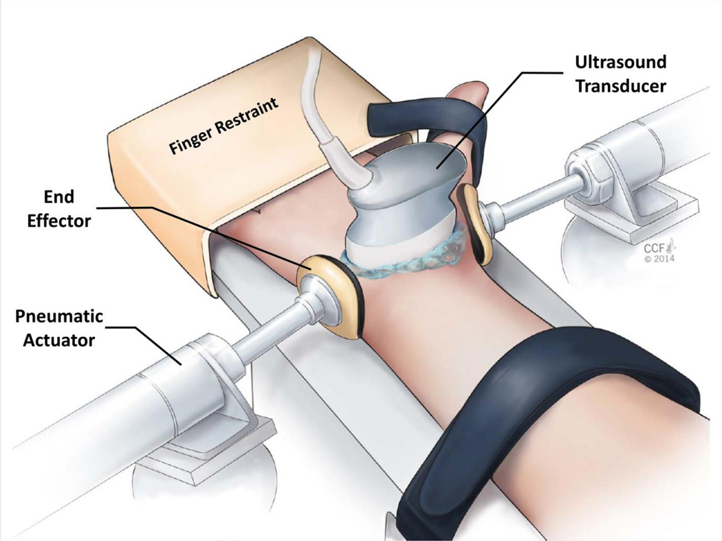
Methods: Ultrasound images of the carpal tunnel and its contents were captured for 11 healthy, female volunteers at the distal tunnel level prior to force application and during force application of 10 and 20N.
Findings: With applied force, the carpal arch width significantly decreased, while the carpal arch height and area significantly increased (P<0.001). The median nerve shape became more rounded as the compressive force magnitude increased, reflected by decreases in the nerve's flattening ratio and increases in its circularity (P<0.001). The applied force also resulted in nerve displacement in the radial-volar direction.
Interpretation: This study demonstrates that noninvasively applying radioulnar compressive force across the wrist may potentially provide relief of median nerve compression to patients suffering from carpal tunnel syndrome.
Keywords: Carpal tunnel syndrome; Compression; Force; Median nerve; Wrist.
Copyright © 2015 Elsevier Ltd. All rights reserved.
Conflict of interest statement
Conflicts of interest: none.
Figures
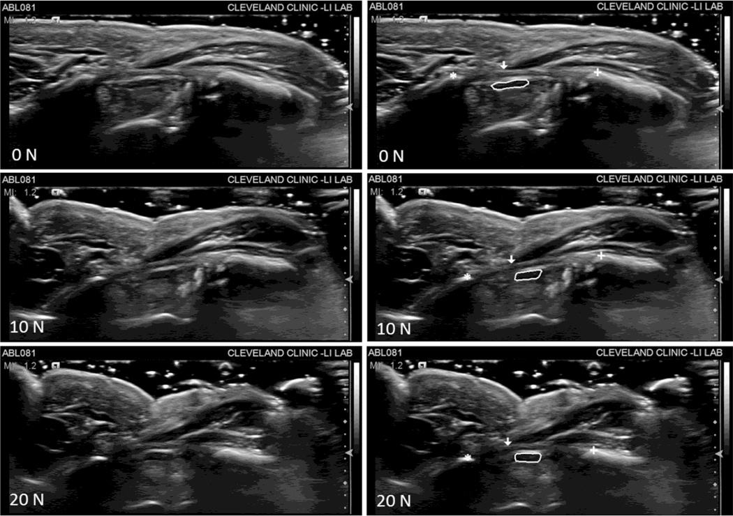
 ), ridge of the trapezium (
 ), thenar muscles ulnar point (
 ), and the median nerve (solid line).
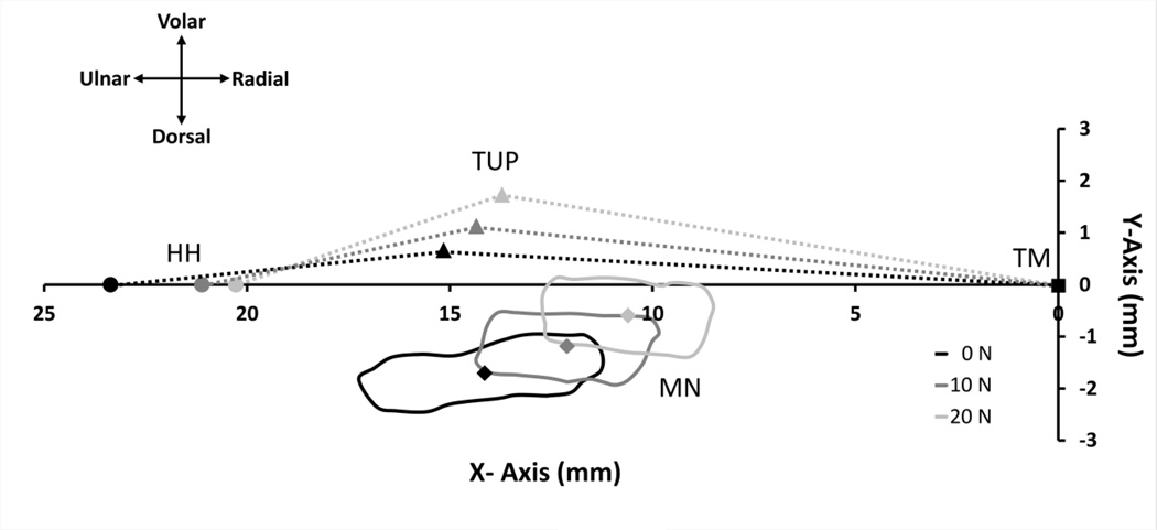
 ), thenar muscles ulnar point (TUP,
  ), and median nerve (MN, solid line) with centroid (
 ) for a representative subject at 0, 10, and 20 N of wrist compression relative to the anatomically defined coordinate system with its origin at the trapezium (TM,
  ). The area beneath the dotted lines bounded by the X-Axis represents the carpal arch area.
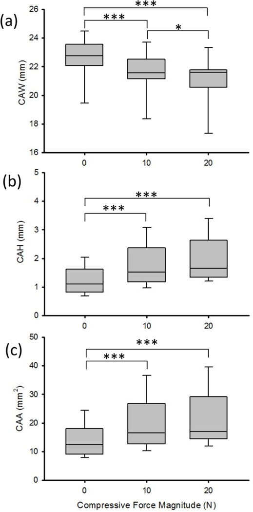
References
-
- Yoshii Y, Ishii T, Tung WL, Sakai S, Amadio PC. Median nerve deformation and displacement in the carpal tunnel during finger motion. J Orthop Res. 2013;31:1876–1880. - PubMed
Publication types
MeSH terms
Grants and funding
LinkOut - more resources
Full Text Sources
Other Literature Sources
Medical
