. 2015 Feb 19;57(4):735-749.
doi: 10.1016/j.molcel.2014.12.037. Epub 2015 Feb 5.

Molecular mechanism and evolution of guanylate kinase regulation by (p)ppGpp

Affiliations

Molecular mechanism and evolution of guanylate kinase regulation by (p)ppGpp

Kuanqing Liu et al. Mol Cell. .

Abstract

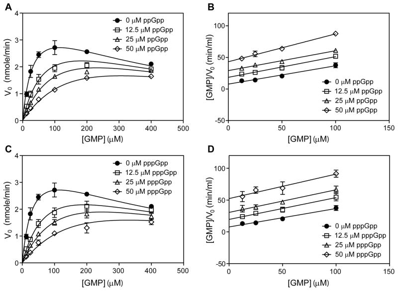
The nucleotide (p)ppGpp mediates bacterial stress responses, but its targets and underlying mechanisms of action vary among bacterial species and remain incompletely understood. Here, we characterize the molecular interaction between (p)ppGpp and guanylate kinase (GMK), revealing the importance of this interaction in adaptation to starvation. Combining structural and kinetic analyses, we show that (p)ppGpp binds the GMK active site and competitively inhibits the enzyme. The (p)ppGpp-GMK interaction prevents the conversion of GMP to GDP, resulting in GMP accumulation upon amino acid downshift. Abolishing this interaction leads to excess (p)ppGpp and defective adaptation to amino acid starvation. A survey of GMKs from phylogenetically diverse bacteria shows that the (p)ppGpp-GMK interaction is conserved in members of Firmicutes, Actinobacteria, and Deinococcus-Thermus, but not in Proteobacteria, where (p)ppGpp regulates RNA polymerase (RNAP). We propose that GMK is an ancestral (p)ppGpp target and RNAP evolved more recently as a direct target in Proteobacteria.


Figures

Figure 1
Figure 1. Inhibition of GMK by pppGpp in selected firmicutes
(A) Proposed alternative mechanisms by which (p)ppGpp regulates transcription in E. coli and B. subtilis to adapt to stresses (e.g. amino acid starvation). (p)ppGpp directly targets RNAP in E. coli, and GMK, HPRT, and IMPDH in B. subtilis. (B) (p)ppGpp inhibits multiple enzymes in the GTP biosynthesis pathways of B. subtilis. The white dots indicate the unoccupied (p)ppGpp binding sites. PRPP: phosphoribosylpyrophosphate; IMP: inosine monophosphate; XMP: xanthosine monophosphate; GMP: guanosine monophosphate; IMPDH: inosine monophosphate dehydrogenase; GMK: guanosine monophosphate kinase (guanylate kinase); HPRT: hypoxanthine-guanine phosphoribosyltransferase. Broken lines indicate multiple steps. (C) Dose-dependent inhibition of firmicute GMKs by pppGpp. Data was fitted into the equation y=100%/[1+(x/IC50)s], where y is the relative enzyme activity, x is the inhibitor concentration, and s is the slope factor. GMK activity was assayed with increasing concentration of pppGpp. Error bars represent standard error of the mean from three replicates. All the tested GMKs contain an N-terminus 6xHis tag, but the tag does not affect the sensitivity of GMK to pppGpp (Figure S1A). Compared to pppGpp, GTP only slightly inhibits firmicute GMKs (Figure S1C). (D) Sequence alignment of GMKs from selected firmicutes using MEGA (Tamura et al., 2011). Residues that interact with pppGpp only and both pppGpp and GMP are highlighted in yellow and light blue, respectively. Domains are assigned as previously described with minor modification (Hible et al., 2005). S. aureus GMK serves as the reference and identical residues are shown as dots. See also Figure S1.
Figure 2
Figure 2. (p)ppGpp competitively inhibits GMK activity
(A and B) ppGpp competitively inhibits S. aureus GMK. Data is fitted into a modified competitive inhibition equation: V0=Vmax[S]/{Km(1+[I]/Ki)+[S](1+[S]/Ki')} to account for substrate (GMP) inhibition (Supplemental Experimental Procedures) (A). Selected data points ([GMP] ≤ 100 μM) are transformed into the Hanes-Woolf equation: [S]/V0=[S]/Vmax+Km/Vmax, to minimize the effect of substrate inhibition on the linear regression (B). (C and D) pppGpp competitively inhibits S. aureus GMK. Data fitting and transformation are described as in (A and B). See also Figure S2.
Figure 3
Figure 3. Structure of pppGpp-bound S. aureus GMK
(A) Omit electron-density map of one selected pppGpp/Mg2+ contoured at 2 σ with the refined ligand structure shown. (B) Apo (green) and pppGpp-bound (blue) GMK are superimposed to indicate the differences between the two structures. (C) GMP (magenta) (El Omari et al., 2006) and pppGpp-bound GMK (blue) are superimposed. The positions of atoms shared between GMP and pppGpp are nearly identical in the two structures. (D) Detailed view of the pppGpp-binding pocket on GMK. Several residues from GMK recognize the base (Ser39, Glu74, Ile104, and Glu105), sugar (Glu103), 5′ triphosphate (Arg43, Arg46, Tyr55, Tyr78, and Tyr83), and 3′ diphosphate (Lys19, and Tyr78) elements of pppGpp. Yellow residues interact with pppGpp only, whereas blue residues interact with both pppGpp and GMP. (E) Summary of kinetic parameters for GMKs. The dissociation constant (Ki) of pppGpp is shown in boldface.
Figure 4
Figure 4. Conservation of regulation of GMK by pppGpp in bacteria
(A) A rooted 16S rRNA phylogenetic tree constructed by the neighbor-joining method using MEGA (Tamura et al., 2011), showing bootstrap values higher than 75. Species are colored based on the sensitivity of their GMKs to pppGpp, and S. cerevisiae (18s rRNA) serves as an outgroup. The length of the radius branches indicates the amount of change (substitutions/site) between a pair of nodes. Inhibition curves of GMKs with pppGpp for selected species in (B) Actinobacteria, (C) Deinococcus-Thermus, (D) α-Proteobacteria, and (E) γ- and β-Proteobacteria, as well as S. cerevisiae. Error bars represent standard error of the mean from three replicates, except for S. cerevisiae for which two replicates were performed. Data fitting is as described in Figure 1C. See also Figure S3 and 4.
Figure 5
Figure 5. E. coli GMK has a small substrate binding pocket that does not fit pppGpp
(A) Isothermal titration calorimetry (ITC) graphs of 50 μM B. subtilis GMK and E. coli GMK titrated with 1 mM pppGpp. (B) Structural comparison between pppGpp-bound S. aureus GMK and GMP-bound E. coli GMK (PDB: 2ANB) (Hible et al., 2005) by aligning the latter to the former (residues 1–124). Molecular docking of pppGpp into S. aureus GMK (C) and E. coli GMK (D), using AutoDock Vina (Trott and Olson, 2009). GMKs (the receptors) were prepared from the pppGpp-bound S. aureus GMK structure (Figure 3) and the GMP-bound E. coli GMK structure (PDB: 2ANB) (Hible et al., 2005), respectively. pppGpp (the ligand) was prepared from the pppGpp-bound S. aureus GMK structure (Figure 3). Docked pppGpp with the highest binding affinity to GMK is shown. (E) Schematic of chimeric GMKs. (F) Characterization of chimeric GMKs constructed in (E) with pppGpp. E. coli and S. aureus GMKs are shown here for comparison. (G) Ser13 and Arg149 of E. coli GMK (black sticks) are likely to interfere with pppGpp binding by steric clashes. The GMP-bound E. coli GMK (PDB: 2ANB) (Hible et al., 2005) was aligned to the pppGpp-bound S. aureus GMK as described in (B). For visualization of the steric clashes (yellow circles), residues 16–26, 38, 40–54, and 128–137 are purposefully hidden in the enlarged insets. See also Figure S5.
Figure 6
Figure 6. Abolishing regulation of GMK by (p)ppGpp leads to defective adaptation to amino acid starvation
(A) Semi-log plot of growth of WT and ecgmk in different nutrient conditions. Error bars (shown as thin lines) represent standard error of the mean from 6 replicates. Schematic of the competition assay (B), and the amino acid downshift and nucleotide detection by LC-MS (C). (D) Percentage of ecgmk in the total population in different nutrient conditions for three continuous growth cycles. Shown here is the geometric average of the two parallel experiments to eliminate the effects of lacZ and error bars represent standard error of the mean from three replicates. Change of GMP (E), GDP (F), and GTP (G) in WT and ecgmk after amino acid downshift. Average of at least two replicates was plotted with standard error of the mean shown. (H) Change of intracellular GTP and GMP in WT during amino acid starvation. Error bars represent the standard error of the mean from at least three replicates. Quantification of intracellular GTP and GMP were described in the Experimental Procedures. (I) (p)ppGpp levels in WT and ecgmk treated with 0.5 mg/ml arginine hydroxamate for 20 minutes. The abundance is defined as PhosphorImager count/OD600. Two replicates for each strain were performed on the same day and samples were loaded onto the same TLC plate so that the PhosphorImager counts are directly comparable. The p-value was calculated using unpaired two-tailed Student’s t-test. See also Figure S6.
Figure 7
Figure 7. Evolution of (p)ppGpp-mediated regulation of GMK and RNAP
(A) Proposed conservation of (p)ppGpp-GMK/RNAP regulation in bacteria. Residues corresponding to the ppGpp-binding residues identified on the E. coli RNA polymerase (β′ and ω subunits) (Ross et al., 2013; Zuo et al., 2013) are aligned from different species. Presence of DksA/DksA-like protein is proposed based on either published work or BLAST (Basic Local Alignment Search Tool; http://blast.ncbi.nlm.nih.gov/Blast.cgi) search using the E. coli DksA as the query. The R. sphaeroides DksA homolog (RSP2654) has been recently characterized and mechanistically resembles the E. coli DksA (Lennon et al., 2014). The conserved DxxDxA motif has been suggested to be an important determinant of DksA functions (Furman et al., 2012) and is utilized here as a criterion together with sequence homology search to determine the presence or absence of DksA-like proteins. Sequence alignment of DksA/DksA-like proteins is summarized in Figure S3B. (B) Phylogenetic tree and inferred evolutionary trajectory of (p)ppGpp-mediated regulation of GMK and RNAP. The phylogenetic tree was constructed as described in Figure 4A, and shown here as a rectangular tree.

Similar articles

Cited by

References

    1. Abranches J, Martinez AR, Kajfasz JK, Chavez V, Garsin DA, Lemos JA. The molecular alarmone (p)ppGpp mediates stress responses, vancomycin tolerance, and virulence in Enterococcus faecalis. J Bacteriol. 2009;191:2248–2256. - PMC - PubMed
    1. Adams PD, Afonine PV, Bunkoczi G, Chen VB, Davis IW, Echols N, Headd JJ, Hung LW, Kapral GJ, Grosse-Kunstleve RW, et al. PHENIX: a comprehensive Python-based system for macromolecular structure solution. Acta Crystallogr Sect D Biol Crystallogr. 2010;66:213–221. - PMC - PubMed
    1. Atkinson GC, Tenson T, Hauryliuk V. The RelA/SpoT homolog (RSH) superfamily: distribution and functional evolution of ppGpp synthetases and hydrolases across the tree of life. PLoS One. 2011;6:e23479. - PMC - PubMed
    1. Bennett HJ, Pearce DM, Glenn S, Taylor CM, Kuhn M, Sonenshein AL, Andrew PW, Roberts IS. Characterization of relA and codY mutants of Listeria monocytogenes: identification of the CodY regulon and its role in virulence. Mol Microbiol. 2007;63:1453–1467. - PubMed
    1. Bittner AN, Kriel A, Wang JD. Lowering GTP level increases survival of amino acid starvation but slows growth rate for Bacillus subtilis cells lacking (p)ppGpp. J Bacteriol. 2014;196:2067–2076. - PMC - PubMed

Publication types

MeSH terms

Associated data

LinkOut - more resources