. 2015 Feb 9:5:8317.
doi: 10.1038/srep08317.

Optogenetic induction of contractile ability in immature C2C12 myotubes

Affiliations

Optogenetic induction of contractile ability in immature C2C12 myotubes

Toshifumi Asano et al. Sci Rep. .

Abstract

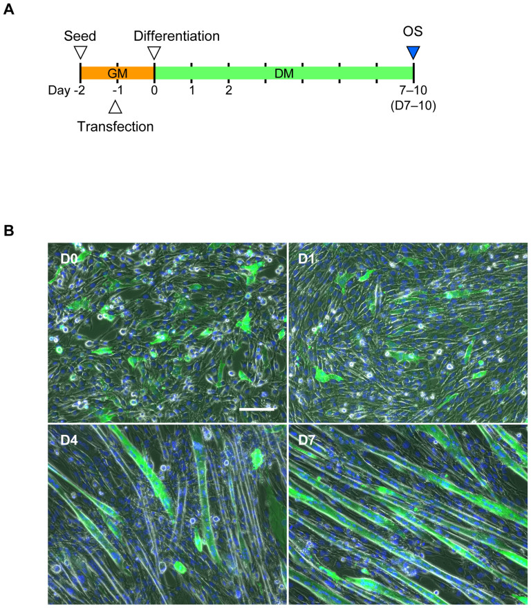
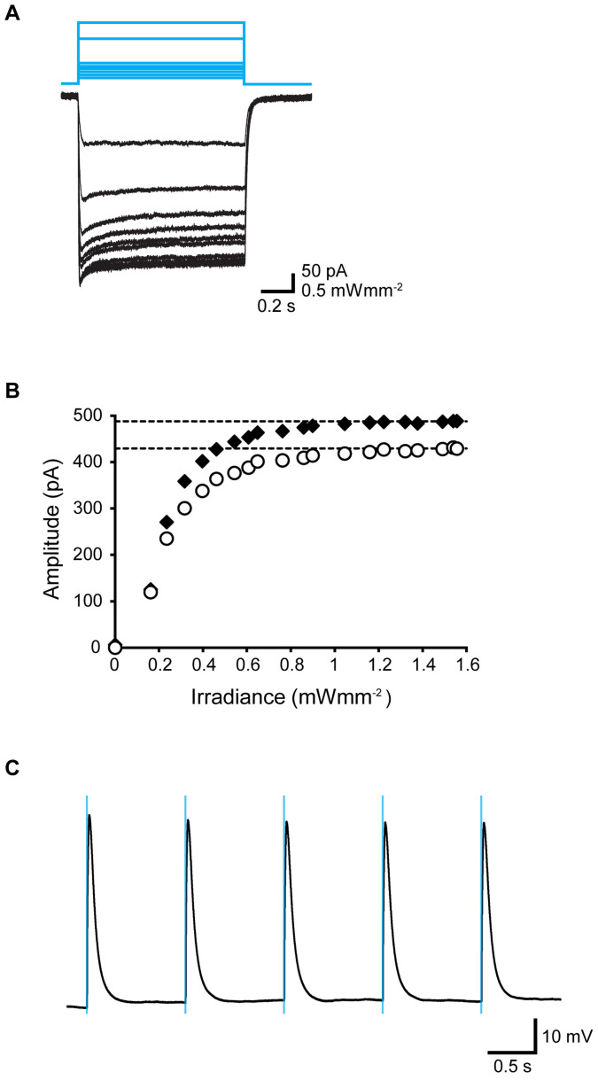
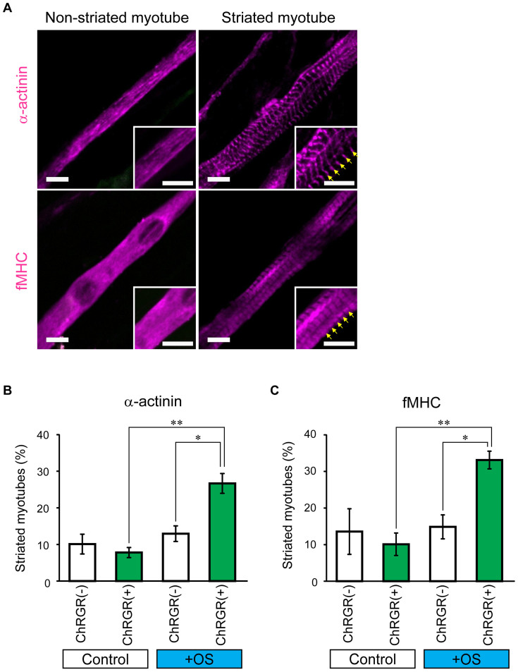
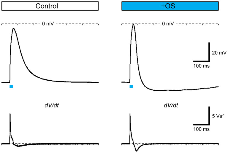
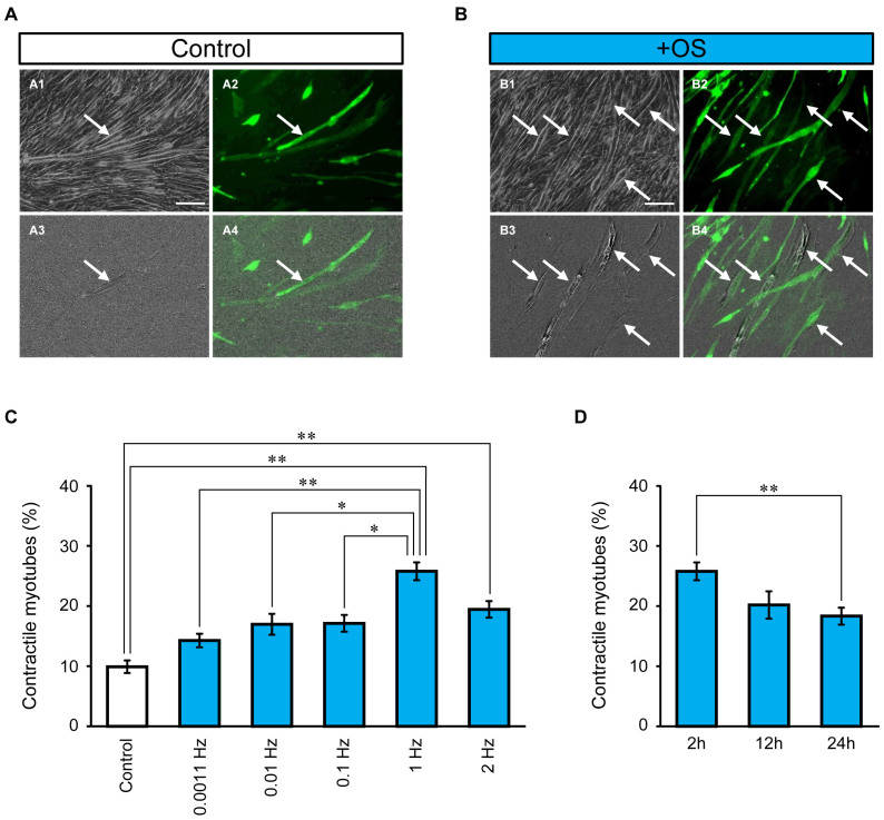
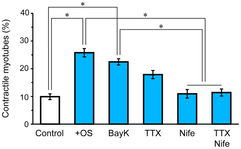
Myoblasts can be differentiated into multinucleated myotubes, which provide a well-established and reproducible muscle cell model for skeletal myogenesis in vitro. However, under conventional differentiation conditions, each myotube rarely exhibits robust contraction as well as sarcomere arrangement. Here, we applied trains of optical stimulation (OS) to C2C12 myotubes, which were genetically engineered to express a channelrhodopsin variant, channelrhodopsin-green receiver (ChRGR), to investigate whether membrane depolarization facilitates the maturation of myotubes. We found that light pulses induced membrane depolarization and evoked action potentials in ChRGR-expressing myotubes. Regular alignments of sarcomeric proteins were patterned periodically after OS training. In contrast, untrained control myotubes rarely exhibited the striated patterns. OS-trained and untrained myotubes also differed in terms of their resting potential. OS training significantly increased the number of contractile myotubes. Treatment with nifedipine during OS training significantly decreased the fraction of contractile myotubes, whereas tetrodotoxin was less effective. These results suggest that oscillations of membrane potential and intracellular Ca(2+) accompanied by OS promoted sarcomere assembly and the development of contractility during the myogenic process. These results also suggest that optogenetic techniques could be used to manipulate the activity-dependent process during myogenic development.


Figures

Figure 1
Figure 1. Generation of photosensitive C2C12 myotubes.
(A) Time schedule for the OS training assay with C2C12 myotubes. One day after seeding (Day −1), cells were transfected with ChRGR-Ve in the growth medium (GM). Twenty-four hours later (Day 0, D0), the medium was replaced with differentiation medium (DM) to promote the formation of multinuclear myotubes. The OS training assay was performed on Days 7–10. (B) Generation of ChRGR-Ve-expressing multinucleated myotubes on day 0 (D0), day 1 (D1), day 4 (D4), and day 7 (D7) after switching to DM. Multinucleated myotubes expressing ChRGR were detected based on Venus-fluorescence (green), and the cell nuclei were stained with Hoechst 33258 (blue). Scale bar, 50 μm.
Figure 2
Figure 2. Optical responses of ChRGR-expressing C2C12 myotubes.
(A) Typical photocurrent traces (black lines) from a photosensitive multinucleated myotube induced by cyan LED light at various intensities (blue lines; 0.16–1.58 mWmm−2). (B) Power dependency of the peak current (filled diamonds) and steady-state current amplitudes (open circles). (C) Rhythmic action potentials generated by repetitive cyan LED flashes (blue lines; duration, 20 ms; frequency, 1 Hz) with the current clamp.
Figure 3
Figure 3. Effects of OS training on sarcomere assembly in ChRGR-expressing C2C12 myotubes.
(A) Images of typical multinucleated myotubes 7 days differentiation induction. Cells were fixed and stained with anti-sarcomeric α-actinin (upper panel) or fMHC (lower panel). The insets show the magnified images. The sarcomeric structures are indicated by arrows. Scale bar, 10 μm. (B) Fractions of myotubes with mature striation patterns. Assembly of α-actinin (from left to right): control ChRGR-negative myotubes without OS training (white column, n = 12 regions), ChRGR-positive myotubes without OS training (green column, n = 12 regions), ChRGR-negative myotubes with OS training (white column, n = 16 regions), and ChRGR-positive myotubes with OS training (green column, n = 16 regions). (C) Similar to (B), but showing the assembly of fMHC in ChRGR-negative (white bar, each, n = 16 regions) and –positive cells (green bar, each, n = 16 regions). Each experiment consists of at least three independent replicates. *, P < 0.05; **, P < 0.01; Kruskal–Wallis test.
Figure 4
Figure 4. Electrical properties of membranes.
Typical action potentials (upper traces) and their time-derivatives (dV/dt data, lower traces) generated by cyan LED flashes (20 ms, indicated by blue lines) in ChRGR-expressing myotubes without (left) or with OS training (right).
Figure 5
Figure 5. Induction of contractile ability.
(A) Image difference extraction analysis of myotubes without OS training: phase-contrast image (A1), ChRGR-Ve-fluorescence image (A2), difference image (A3), and merged image of A2 and A3 (A4). Contracted myotubes are indicated by arrows. (B) Myotubes after OS training. Scale bar, 100 μm. (C) Effects of OS protocols on the fraction of contractile fluorescent myotubes divided by the total fluorescent myotubes (from left to right): control without OS (n = 24 regions), 0.0011 Hz (n = 20 regions), 0.01 Hz (n = 20 regions), 0.1 Hz (n = 28 regions), 1 Hz (n = 32 regions), and 2 Hz (n = 16 regions). (D) Fraction of contractile myotubes with OS protocols at 1 Hz for (from left to right): 2 h (n = 32 regions), 12 h (n = 12 regions), and 24 h (n = 16 regions). Each experiment consists of at least four independent replicates. *, P < 0.05; **, P < 0.01; Kruskal–Wallis test.
Figure 6
Figure 6. Effects of pharmacological treatments on the induction of the contractile ability.
From left to right: without OS training as a reference (n = 24 regions), OS training without any treatment (n = 32 regions), BayK (n = 20 regions), TTX (n = 20 regions), Nife (n = 24 regions), and TTX and Nife (n = 16 regions) in ChRGR-expressing myotubes. Each experiment consists of at least four independent replicates. *, P < 0.01; Kruskal–Wallis test.

References

    1. Booth F. W., Chakravarthy M. V., Gordon S. E. & Spangenburg E. E. Waging war on physical inactivity: using modern molecular ammunition against an ancient enemy. J. Appl. Physiol. 93, 3–30 (2002). - PubMed
    1. Handschin C. & Spiegelman B. M. The role of exercise and PGC1α in inflammation and chronic disease. Nature 454, 463–469 (2008). - PMC - PubMed
    1. Flück M. Functional, structural and molecular plasticity of mammalian skeletal muscle in response to exercise stimuli. J. Exp. Biol. 209, 2239–2248 (2006). - PubMed
    1. Sketelj J. et al. Specific impulse patterns regulate acetylcholinesterase activity in skeletal muscles of rats and rabbits. J. Neurosci. Res. 47, 49–57 (1997). - PubMed
    1. Freud-Silverberg M. & Shainberg A. Electric stimulation regulates the level of Ca-channels in chick muscle culture. Neurosci. Lett. 151, 104–106 (1993). - PubMed

Publication types