. 2015 Mar 3;23(3):461-471.
doi: 10.1016/j.str.2014.12.016. Epub 2015 Feb 5.

Structure of the vacuolar H+-ATPase rotary motor reveals new mechanistic insights

Affiliations

Structure of the vacuolar H+-ATPase rotary motor reveals new mechanistic insights

Shaun Rawson et al. Structure. .

Abstract

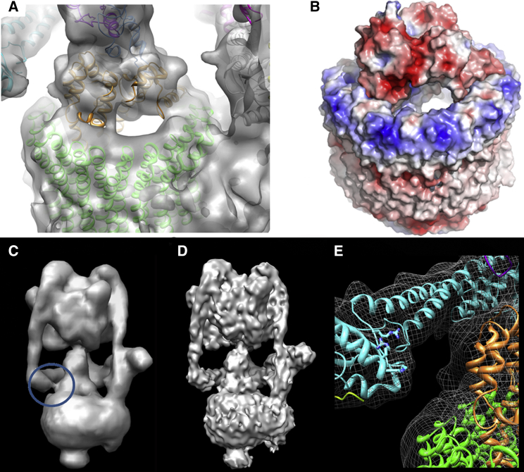
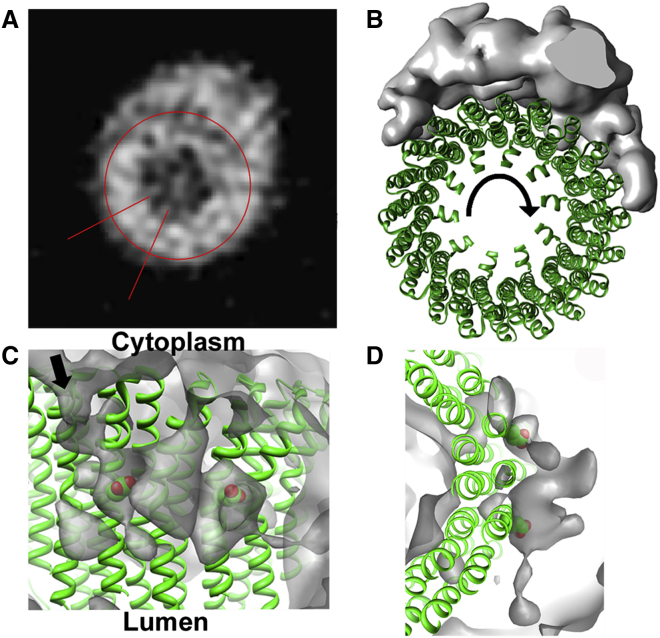
Vacuolar H(+)-ATPases are multisubunit complexes that operate with rotary mechanics and are essential for membrane proton transport throughout eukaryotes. Here we report a ∼ 1 nm resolution reconstruction of a V-ATPase in a different conformational state from that previously reported for a lower-resolution yeast model. The stator network of the V-ATPase (and by implication that of other rotary ATPases) does not change conformation in different catalytic states, and hence must be relatively rigid. We also demonstrate that a conserved bearing in the catalytic domain is electrostatic, contributing to the extraordinarily high efficiency of rotary ATPases. Analysis of the rotor axle/membrane pump interface suggests how rotary ATPases accommodate different c ring stoichiometries while maintaining high efficiency. The model provides evidence for a half channel in the proton pump, supporting theoretical models of ion translocation. Our refined model therefore provides new insights into the structure and mechanics of the V-ATPases.


Figures

None
Graphical abstract
Figure 1
Figure 1
3D Reconstruction of the Manduca sexta V-ATPase at ∼1 nm Resolution (A and B) Surface views rotated by 120°, note the open AB site faces the viewer in (B). S1–S3 are stator filaments comprising E and G subunits. Scale bar, 60 Å. (C) Molecular models of M. sexta subunits fitted into the reconstruction, based on crystal structures of homologs from Saccharomyces cerevisiae V-ATPase or bacterial A-ATPases. See also Movie S1. (D) Section through the electron density map of the V1 midsection, with the AB active sites indicated by a green triangle, open site by an open triangle, and the noncatalytic AB interfaces by red triangles. (E) Representative electron density in the V1 domain, taken from the square section in (A), around the DELSEED-related region showing the quality of crystal structure fitting.
Figure 2
Figure 2
Stator Connections in the V-ATPase (A) Transparent surface view of the V1 domain of M. sexta V-ATPase with subunits docked, showing the differences between the “open” and “closed” AB domains. Subunits are colored as in Figure 1C. (B) Superposition of stator filaments 1, 2, and 3 of M. sexta (mesh) and yeast (green surface) V-ATPase based on a global alignment, showing the very similar EG conformations despite differences in the AB catalytic state. See also Figure S1. (C) Comparisons of the interface for each stator, with electrostatic surface shown above and cartoon representation below. The EG stator, C subunit, H subunit, and a subunit are yellow, cyan, magenta, and gray, respectively.
Figure 3
Figure 3
Motor-Axle Interactions in V1 (A) Vertical section through V1, with the EM map shown in mesh format and models of subunits A (red), B (blue), and D (cyan) fitted. Distinctive contacts can be seen at the lever (L) and bearing (B) regions. (B) Slice-through of the lever region, and (C) bearing region showing the close packing against the P-A/G-D/E-X-G-Y/F-P (subunit A) and P-G/S-R-R/K-G-Y/F-P (subunit B) loops. (D) Comparison of the Enterococcus hirae A3B3DF crystal structure (red) and M. sexta A3B3DF model (blue). (E and F) Superposition of the open (cyan), loose (green), and tightly bound (magenta) A (E) and B (F) subunits. Greatest differences are at the base region corresponding to the lever arm domain (L) involved in torque generation. Little change is observed within the bearing region (B). See also Movie S2. (G) Sequence conservation in V1, calculated in Consurf (Goldenberg et al., 2009). The continuum ranges from pink (highly conserved) to black (no conservation). Strongest sequence identity is found within the bearing region. (H) Electrostatic surfaces at the bearing region of M. sexta V-ATPase shown on a scale of −5.0 (red) to 5.0 (blue). (I) Superposition of the conserved loop region for subunits A (red) and B (blue) from M. sexta V-ATPase, A (yellow) and B (green) from Enterococcus hirae A-ATPase (Arai et al., 2013), α (cyan) and β (wheat) from bovine mitochondrial F-ATPase (Bowler et al., 2007), and FliI (magenta) from the flagella motor (Imada et al., 2007). The position of the rotor axle subunit D is shown as a gray helix. In (B), (C), and (H) the V-ATPase is viewed from the luminal side and rotation of the axle will be counterclockwise.
Figure 4
Figure 4
Rotor Coupling in the V-ATPase (A) M. sexta map around the c ring/subunit d connection. Significant cavities between the subunits are evident. (B) Electrostatics of the c ring/d subunit interface shows the strong net negative charge of d complementing the positive charge of the c ring. Electrostatics were calculated and scaled as detailed in Figure 3. Note that both (A) and (B) are in approximately the same orientation. (C and D) Apparent contact between subunits C and d in the M. sexta reconstructions at 11 Å (C) and 9.4 Å (D). The “linker” region is circled. (E) Cartoon of subunits C (cyan), d (orange), and the c ring (green). Residues making a positive surface are in stick format and are 99% (Arg248), 87% (Lys244), and 95% (Arg241) conserved at that position, based on a 200-sequence comparison using Consurf (Goldenberg et al., 2009).
Figure 5
Figure 5
Organization of Vo (A) Section through the Vo region of the map. The red circle shows the approximate boundary of the c ring and lines delineate a four-helix bundle of one c subunit. (B) Segmentation of the map showing the extent of subunit a around the c ring (represented by the NtpK 10-mer structure (PDB ID 2BL2; green). Arrow donates direction of rotation. (C and D) Region of low-density “cavities,” enclosed within the gray surface, at the c ring (green)/subunit a interface consistent with a proton-accessible “half channel” (arrow). The conserved glutamate in helix 4 of subunit c playing a key role in proton transfer is shown in stick format. This occurs at the same depth in the membrane as the low-density feature, which is at a position appropriate for the previously hypothetical “proton half channel,” discussed in the text. Views are parallel to the plane of the membrane (C) and from the luminal side (D).
Figure 6
Figure 6
Deglycosylation Analysis of M. sexta V-ATPase (A) Image sums of the full data sets from V-ATPase (i) in the native untreated state (V-ATPasen), (ii) treated as for deglycosylated enzyme but lacking PNGase F (V-ATPasec), (iii) and (iv) treated with PNGase F at 17°C (V-ATPased17) or 30°C (V-ATPased30), respectively. (B) Image sum and masks used. (C and D) Comparable classes from the control (left) and deglycosylated sample (right) of two different side views. (E) Percentage of particles displaying density at the base of Vo in the native, control, and PNGase-incubated V-ATPase (with treatments at 17°C and 30°C). Each data set was processed with the three different masks shown in (B) to check for processing artifacts.
Figure 7
Figure 7
Data Processing of M. sexta V-ATPase (A) Representative cryo-EM micrograph of M. sexta V-ATPase on a 10 nm thick carbon support film. (B) Reference-free classes generated in RELION; note the clarity of the stator connections. (C) FSC plot for the resulting RELION reconstruction.

References

    1. Abrahams J.P., Leslie A.G., Lutter R., Walker J.E. Structure at 2.8 Å resolution of F1-ATPase from bovine heart mitochondria. Nature. 1994;370:621–628. - PubMed
    1. Angevine C.M., Herold K.A.G., Vincent O.D., Fillingame R.H. Aqueous access pathways in ATP synthase subunit A—reactivity of cysteine substituted into transmembrane helices 1, 3, and 5. J. Biol. Chem. 2007;282:9001–9007. - PubMed
    1. Arai S., Saijo S., Suzuki K., Mizutani K., Kakinuma Y., Ishizuka-Katsura Y., Ohsawa N., Terada T., Shirouzu M., Yokoyama S. Rotation mechanism of Enterococcus hirae V1-ATPase based on asymmetric crystal structures. Nature. 2013;493:703–707. - PubMed
    1. Beltran C., Nelson N. The membrane sector of vacuolar H(+)-ATPase by itself is impermeable to protons. Acta Physiol. Scand. Suppl. 1992;607:41–47. - PubMed
    1. Benlekbir S., Bueler S.A., Rubinstein J.L. Structure of the vacuolar-type ATPase from Saccharomyces cerevisiae at 11Å resolution. Nat. Struct. Mol. Biol. 2012;19:1356–1362. - PubMed

Publication types

Substances

LinkOut - more resources