Review
. 2015 Jun;72(11):2041-59.
doi: 10.1007/s00018-015-1847-9. Epub 2015 Feb 7.

The PRMT5 arginine methyltransferase: many roles in development, cancer and beyond

Affiliations
Review

The PRMT5 arginine methyltransferase: many roles in development, cancer and beyond

Nicole Stopa et al. Cell Mol Life Sci. 2015 Jun.

Abstract

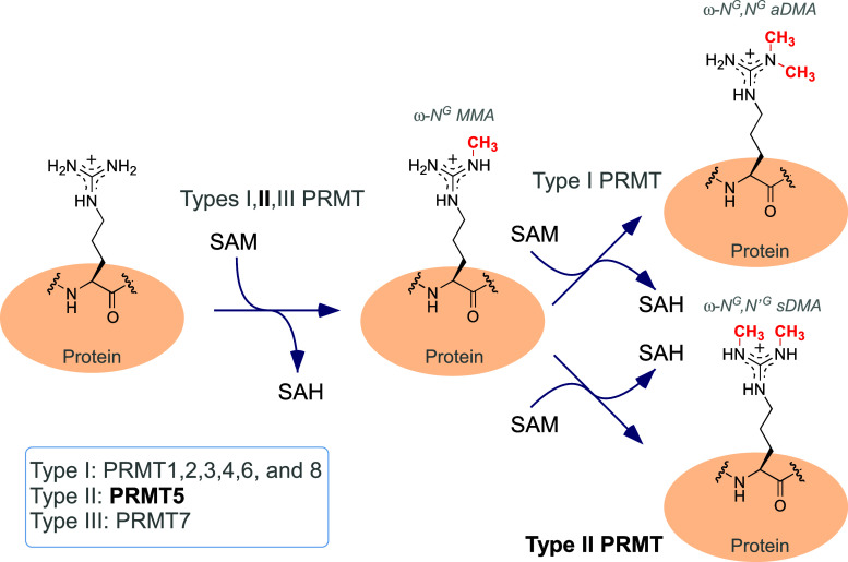
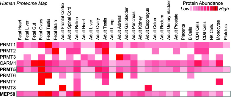
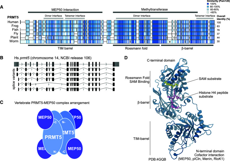
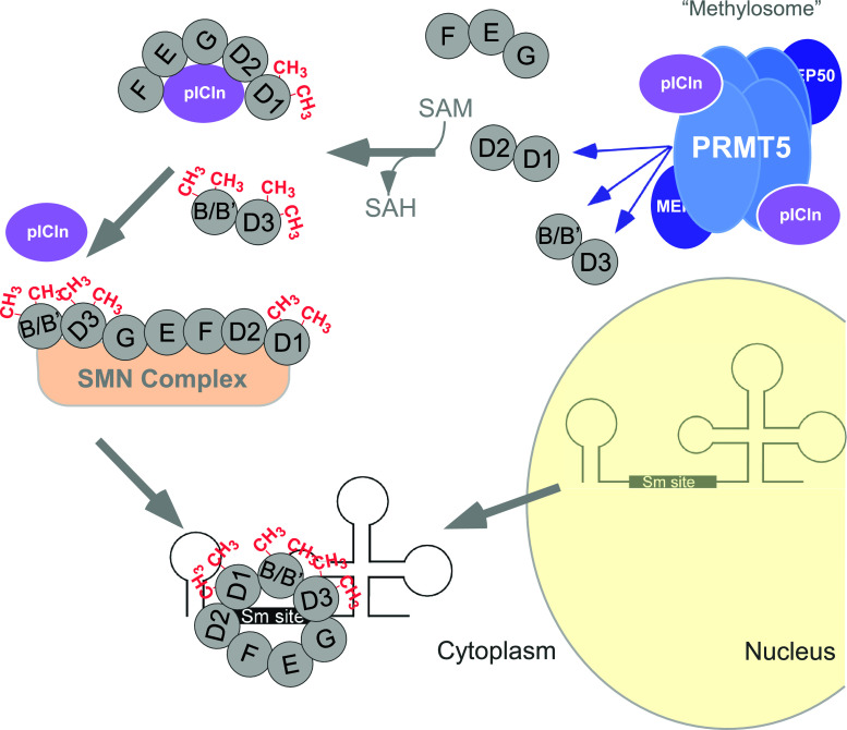
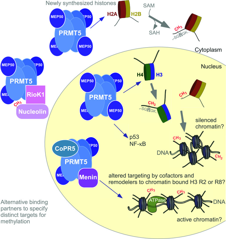
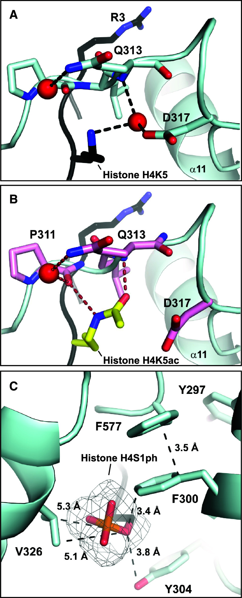
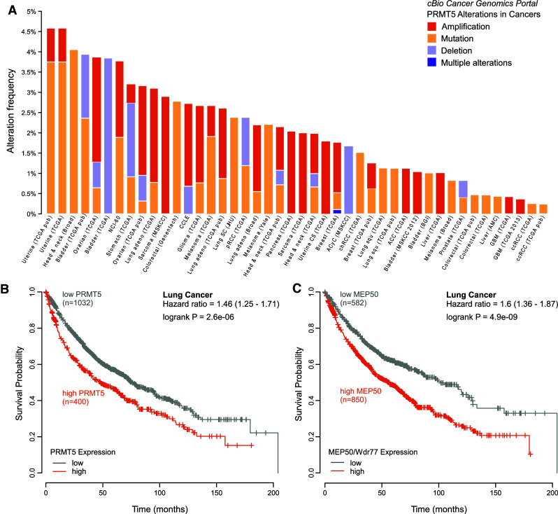
Post-translational arginine methylation is responsible for regulation of many biological processes. The protein arginine methyltransferase 5 (PRMT5, also known as Hsl7, Jbp1, Skb1, Capsuleen, or Dart5) is the major enzyme responsible for mono- and symmetric dimethylation of arginine. An expanding literature demonstrates its critical biological function in a wide range of cellular processes. Histone and other protein methylation by PRMT5 regulate genome organization, transcription, stem cells, primordial germ cells, differentiation, the cell cycle, and spliceosome assembly. Metazoan PRMT5 is found in complex with the WD-repeat protein MEP50 (also known as Wdr77, androgen receptor coactivator p44, or Valois). PRMT5 also directly associates with a range of other protein factors, including pICln, Menin, CoPR5 and RioK1 that may alter its subcellular localization and protein substrate selection. Protein substrate and PRMT5-MEP50 post-translation modifications induce crosstalk to regulate PRMT5 activity. Crystal structures of C. elegans PRMT5 and human and frog PRMT5-MEP50 complexes provide substantial insight into the mechanisms of substrate recognition and procession to dimethylation. Enzymological studies of PRMT5 have uncovered compelling insights essential for future development of specific PRMT5 inhibitors. In addition, newly accumulating evidence implicates PRMT5 and MEP50 expression levels and their methyltransferase activity in cancer tumorigenesis, and, significantly, as markers of poor clinical outcome, marking them as potential oncogenes. Here, we review the substantial new literature on PRMT5 and its partners to highlight the significance of understanding this essential enzyme in health and disease.


Figures

Fig. 1
Fig. 1
Arginine methylation states catalyzed by the family of protein arginine methyltransferases (PRMTs). The guanidinium side chain of arginine residues in proteins is positively charged. It can accept a monomethyl addition, catalyzed by the family of Type I, II, and III PRMTs through transfer from the S-adenosylmethionine (SAM or AdoMet) cosubstrate, resulting in a ω-N G monomethylated arginine (MMA) and S-adenosylhomocysteine (SAH). Type I PRMTs, comprising the majority of PRMT enzymes, can further catalyze the ω-N G monomethylation to ω-N GN G asymmetric dimethylarginine (aDMA), consuming SAM and producing SAH. PRMT5, a Type II enzyme, catalyzes the ω-N G monomethylation to ω-N G, NG asymmetric dimethylarginine (sDMA), also consuming SAM and producing SAH. Type III enzymes are incapable of processing to dimethylation. Methylation does not alter the positive charge on the arginine guanidinium side chain
Fig. 2
Fig. 2
PRMT5 and MEP50 are broadly expressed in somatic and embryonic tissues. The human proteome map, analyzed by total proteome mass spectrometry (http://www.humanproteomemap.org [7]), was queried for the PRMT family of proteins which showed that they are distinctly expressed in a range of human tissues and cells. The relative protein abundances for the PRMT1-8 (CARM1 is the name for PRMT4) are shown in a heatmap, with white representing low protein abundance and dark red representing higher abundance, with a ten-step range indicated in the legend. PRMT5 is bolded and boxed, as is its MEP50 cofactor. Note that PRMT5 and MEP50 are most highly expressed in fetal tissue and that their expression patterns are quite similar
Fig. 3
Fig. 3
PRMT5 domain organization and structure are evolutionarily conserved. a A range of PRMT5 protein sequences across eukaryotic species [Homo sapiens (human), Xenopus laevis (frog), Danio rerio (fish), Drosophila melanogaster (fly), Arabidopsis thaliana (plant), Caenorhabditis elegans (worm)] was aligned using the MAFFT algorithm and the Pam120 similarity index and represented in a heatmap from white (<60 % similarity) to dark blue (100 % similarity). Alignment gaps are indicated by a line, and overall identity is shown on the right. The major domains and interfaces are indicated above and below the sequences. Asterisk indicates sequence insertion in C. elegans PRMT5 that prohibits tetramerization. b The human prmt5 gene has multiple splice variants, as shown from the NCBI human genome sequence. All the variants are in the N-terminal domain of the encoded protein. c Subunit arrangement of the hetero-octameric PRMT5–MEP50 structure shown in cartoon form, with the head-to-tail N-terminal and C-terminal PRMT5 arrangement shown by “N-” and “-C”. d Ribbon diagram of a monomer of human PRMT5 (PDB:4GQB) with the domains and substrate-binding sites as indicated
Fig. 4
Fig. 4
PRMT5 methylation and regulation of the spliceosome. A cartoon representation of the function of PRMT5 methylation of splicing proteins in the cytoplasm. Methylated substrates are represented with a red “–CH3”. PRMT5, in complex with MEP50 and pICln, form the methylosome that targets spliceosomal subunits for methylation. pICln then chaperones the subunits to the SMN complex, resulting in proper targeting of RNAs to be spliced
Fig. 5
Fig. 5
PRMT5 is targeted to multiple histone and nuclear targets by cofactors. A cartoon representation of the function of PRMT5 methylation of nuclear proteins (nucleus represented by pale yellow). Methylated substrates are represented with a red “–CH3”. Histones, the protein component of chromatin, are synthesized and then transported to the nucleus. PRMT5–MEP50 targets newly synthesized histone H2A in the cytoplasm and may target soluble H4 in the nucleus (both H2A and H4 are methylated on R3 in the sequence N-SGRGK… as shown in the cartoon), as well as transcription factors such as p53 and NF-κB. PRMT5-methylated H2A and H4 are then deposited into chromatin (DNA wrapped around histone proteins, with histone N-terminal tails indicated in the cartoon). Alternative binding partners for PRMT5 (RioK1 in the cytoplasm, CoPR5 and Menin in the nucleus) may displace one or more MEP50 molecules and alter the targeting of PRMT5 toward substrates as shown, including histone H3 on R2 or R8 in the sequence N-ARTKQTARKST…
Fig. 6
Fig. 6
Structural basis for modification crosstalk regulation of PRMT5 activity. The crystal structure of PRMT5–MEP50 complexed with H4 (1–8) tail peptide (PDB:4GQB) provided insight into activity crosstalk by other histone PTMs. a The histone H4 Lys 5 (H4K5, black stick) interacts with PRMT5 through a hydrogen bond between a structural water molecule (red ball) and its ε-NH2. b Modeled interactions between an acetylated histone H4 Lys 5 (H4K5ac, yellow stick) within the HsPRMT5 active site. The oxygen-carbonyl occupies the position of the structural water molecule shown in a. Acetylation of the peptide at the K5 position increases the enzyme/substrate affinity through enhanced hydrogen bonding. c Modeled potential interactions between a phosphorylated histone H4 Ser 1 (H4S1ph) and the enzyme. The potential occupied space of the phosphorylated residue is shown in mesh, and may either sterically block histone peptide interaction, electrostatically repel PRMT5 Y304 in an active site pH-dependent fashion, or alternatively enhance interaction with enzyme and reduce turnover
Fig. 7
Fig. 7
PRMT5 is altered in a range of cancers and its expression is correlated with poor prognosis. a The alteration frequency of prmt5 gene amplification, mutation, and deletions in a wide range of human cancers cataloged in The Cancer Genome Atlas (TCGA, accessed through the cBio Cancer Genomics Portal; http://www.cbioportal.org) was plotted in a histogram, ranging up to 4.5 % alteration in uterine cancer. This analysis did not include increased gene expression or protein abundance. b A Kaplan–Meier survival probability plot for high (orange) versus low (gray) prmt5 gene expression/mRNA level for lung cancer is shown, with high prmt5 expression resulting in a ~1.5-fold worse survival (hazard ratio) at very high significance. c A Kaplan–Meier survival probability plot for high (orange) versus low (gray) mep50 gene expression/mRNA level for lung cancer is shown, with high prmt5 expression resulting in a ~1.6-fold worse survival (hazard ratio) at very high significance. Survival data obtained from http://www.kmplot.com

References

    1. Di Lorenzo A, Bedford MT. Histone arginine methylation. FEBS Lett. 2011;585(13):2024–2031. doi: 10.1016/j.febslet.2010.11.010. - DOI - PMC - PubMed
    1. Niewmierzycka A, Clarke S. S-Adenosylmethionine-dependent methylation in Saccharomyces cerevisiae. Identification of a novel protein arginine methyltransferase. J Biol Chem. 1999;274(2):814–824. doi: 10.1074/jbc.274.2.814. - DOI - PubMed
    1. Bedford MT, Clarke SG. Protein arginine methylation in mammals: who, what, and why. Mol Cell. 2009;33(1):1–13. doi: 10.1016/j.molcel.2008.12.013. - DOI - PMC - PubMed
    1. Migliori V, Muller J, Phalke S, Low D, Bezzi M, Mok WC, Sahu SK, Gunaratne J, Capasso P, Bassi C, Cecatiello V, De Marco A, Blackstock W, Kuznetsov V, Amati B, Mapelli M, Guccione E. Symmetric dimethylation of H3R2 is a newly identified histone mark that supports euchromatin maintenance. Nat Struct Mol Biol. 2012;19(2):136–144. doi: 10.1038/nsmb.2209. - DOI - PubMed
    1. Zurita-Lopez CI, Sandberg T, Kelly R, Clarke SG. Human protein arginine methyltransferase 7 (PRMT7) is a type III enzyme forming omega-NG-monomethylated arginine residues. J Biol Chem. 2012;287(11):7859–7870. doi: 10.1074/jbc.M111.336271. - DOI - PMC - PubMed

Publication types

MeSH terms

LinkOut - more resources