. 2015 Apr;81(8):2696-705.
doi: 10.1128/AEM.00096-15. Epub 2015 Feb 6.

Targeting Enterococcus faecalis biofilms with phage therapy

Affiliations

Targeting Enterococcus faecalis biofilms with phage therapy

Leron Khalifa et al. Appl Environ Microbiol. 2015 Apr.

Abstract

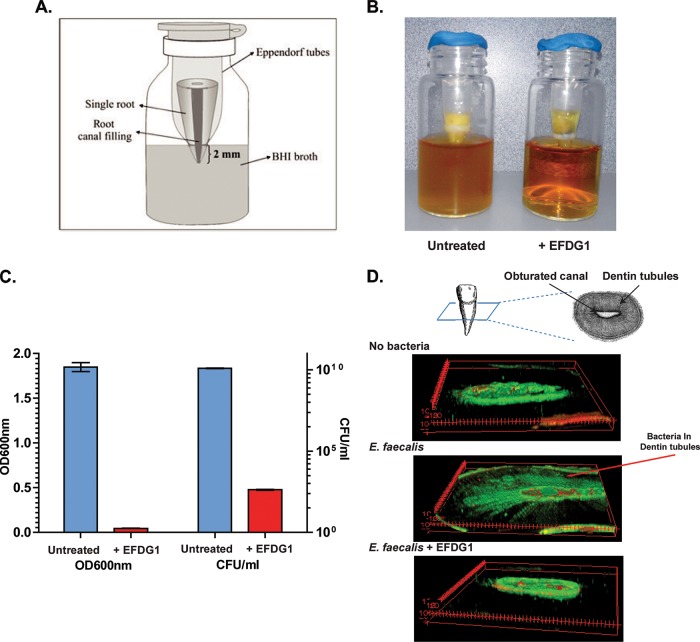
Phage therapy has been proven to be more effective, in some cases, than conventional antibiotics, especially regarding multidrug-resistant biofilm infections. The objective here was to isolate an anti-Enterococcus faecalis bacteriophage and to evaluate its efficacy against planktonic and biofilm cultures. E. faecalis is an important pathogen found in many infections, including endocarditis and persistent infections associated with root canal treatment failure. The difficulty in E. faecalis treatment has been attributed to the lack of anti-infective strategies to eradicate its biofilm and to the frequent emergence of multidrug-resistant strains. To this end, an anti-E. faecalis and E. faecium phage, termed EFDG1, was isolated from sewage effluents. The phage was visualized by electron microscopy. EFDG1 coding sequences and phylogeny were determined by whole genome sequencing (GenBank accession number KP339049), revealing it belongs to the Spounavirinae subfamily of the Myoviridae phages, which includes promising candidates for therapy against Gram-positive pathogens. This analysis also showed that the EFDG1 genome does not contain apparent harmful genes. EFDG1 antibacterial efficacy was evaluated in vitro against planktonic and biofilm cultures, showing effective lytic activity against various E. faecalis and E. faecium isolates, regardless of their antibiotic resistance profile. In addition, EFDG1 efficiently prevented ex vivo E. faecalis root canal infection. These findings suggest that phage therapy using EFDG1 might be efficacious to prevent E. faecalis infection after root canal treatment.


Figures

FIG 1
FIG 1
EFDG1 is an efficient lytic phage that infects E. faecalis. (A) Clear plaques of EFDG1 grown on an E. faecalis lawn. (B) E. faecalis overnight culture was diluted to 1:1,000 in the absence or presence of EFDG1 phages (MOI = 10−4), followed by incubation for 24 h. Total clearance is observed in the treated culture. (C) EFDG1 kills logarithmic E. faecalis in a dose-dependent manner, with an MOI as low as 10−4. Bacterial cultures were grown as in panel B, and the turbidity was measured. (D) EFDG1 effectively reduces stationary cultures of E. faecalis at an MOI as low as 10−7. The results presented in panels B and C are the averages of six independent wells. (E) Validation of the killing by CFU count of E. faecalis bacteria after 24 h (logarithmic, left panel) and 120 h (stationary, right panel) with or without treatment by EFDG1 at MOIs of 10−4 and 10−7, respectively. Bars represent the average of triplicates, and error bars denote the standard deviations.
FIG 2
FIG 2
EFDG1 significantly reduces E. faecalis biofilms. EFDG1 was added to a 2-week-old biofilm of E. faecalis. (A) A confocal 3D image demonstrates that the phage reduces the biofilm almost completely. (B) Quantitative representation of bacteria number within the biofilm layer, as detected by confocal microscopy. These results were also validated, and the kinetics of lysis were determined by crystal violet (CV) staining (C) and a CFU count (D). Tests were performed in triplicates, and all tests yielded significant difference between treated and control groups (P < 0.01). Error bars denote the standard deviations.
FIG 3
FIG 3
Characterization of EFDG1. (A) TEM of EFDG1 depicting a hexagonal head diameter of 98.71 ± 8.88 nm and a tail length118.05 ± 6.87 nm. (B to D) EFDG1 belongs to the Spounavirinae subfamily of the Myoviridae phages. (B) Schematic representation of the EFDG1 DNA sequence and putative genes (green arrows). Red squares denote repeat sequences. The graphs show the GC (blue) and AT (green) content. (C) Phylogenetic tree of EFDG1 (in red) in relation to the genomes of fully sequenced Spounavirinae phages. The name of the phages and their accession numbers in the NCBI database are denoted. (D) Comparison of the EFDG1 genome and its closest related phage, phiEF24c, using the Mauve plugin of Geneious 7.5.1. Similarly colored boxes indicate similar regions.
FIG 4
FIG 4
EFDG1 protects root canals from infection. (A) Schematic depicting the ex vivo root canal treatment model. One-rooted human teeth were subjected to endodontic treatment, including standard cleaning, shaping, and filling. Bacterial contamination was performed before, during, and after the root canal treatment. The test group included phage irrigation in addition to the standard procedure. (B) Photographic depiction of the root irrigated with EFDG1 phage showing clear broth in the lower chamber (48 h), indicating no bacterial outgrowth (right), and control root subjected to the standard protocol, demonstrating broth turbidity (left). (C) OD600 (left axis) and number of live bacteria by CFU count (CFU/ml, right axis) in the lower chamber. Viable E. faecalis counts depicted were reduced by at least 8 logs following phage irrigation. All experiments were repeated at least three times independently, and error bars denote standard deviations. (D) Confocal fluorescence microscopy images of a horizontal root section of the phage treated tooth depicting low numbers of stained bacteria compared to the control, where stained live and dead bacteria are depicted in the dentinal tubules surrounding the root canal (red arrow). Note that the root canal is stained nonspecifically, even in the absence of bacteria.

References

    1. Dahl A, Bruun NE. 2013. Enterococcus faecalis infective endocarditis: focus on clinical aspects. Expert Rev Cardiovasc Ther 11:1247–1257. doi:10.1586/14779072.2013.832482. - DOI - PubMed
    1. Stuart CH, Schwartz SA, Beeson TJ, Owatz CB. 2006. Enterococcus faecalis: its role in root canal treatment failure and current concepts in retreatment. J Endod 32:93–98. doi:10.1016/j.joen.2005.10.049. - DOI - PubMed
    1. Tebruegge M, Pantazidou A, Clifford V, Gonis G, Ritz N, Connell T, Curtis N. 2011. The age-related risk of coexisting meningitis in children with urinary tract infection. PLoS One 6:e26576. doi:10.1371/journal.pone.0026576. - DOI - PMC - PubMed
    1. Koch S, Hufnagel M, Theilacker C, Huebner J. 2004. Enterococcal infections: host response, therapeutic, and prophylactic possibilities. Vaccine 22:822–830. doi:10.1016/j.vaccine.2003.11.027. - DOI - PubMed
    1. Molander A, Reit C, Dahlen G, Kvist T. 1998. Microbiological status of root-filled teeth with apical periodontitis. Int Endod J 31:1–7. doi:10.1046/j.1365-2591.1998.t01-1-00111.x. - DOI - PubMed

Publication types

MeSH terms

Associated data

LinkOut - more resources