Review
. 2015 Feb 6;7(9):a020370.
doi: 10.1101/cshperspect.a020370.

Astrocytes Control Synapse Formation, Function, and Elimination

Affiliations
Review

Astrocytes Control Synapse Formation, Function, and Elimination

Won-Suk Chung et al. Cold Spring Harb Perspect Biol. .

Abstract

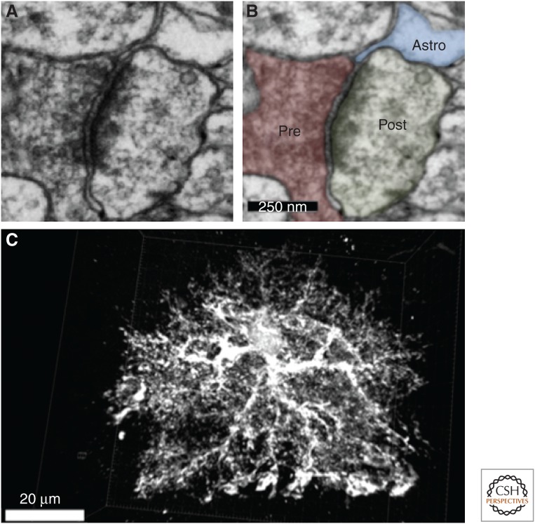
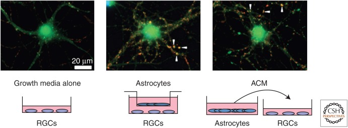
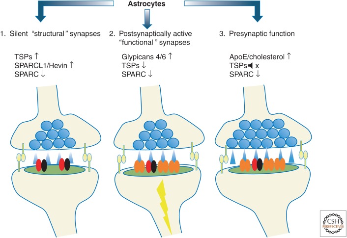
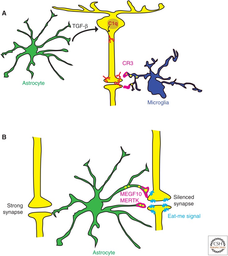
Astrocytes, through their close associations with synapses, can monitor and alter synaptic function, thus actively controlling synaptic transmission in the adult brain. Besides their important role at adult synapses, in the last three decades a number of critical findings have highlighted the importance of astrocytes in the establishment of synaptic connectivity in the developing brain. In this article, we will review the key findings on astrocytic control of synapse formation, function, and elimination. First, we will summarize our current structural and functional understanding of astrocytes at the synapse. Then, we will discuss the cellular and molecular mechanisms through which developing and mature astrocytes instruct the formation, maturation, and refinement of synapses. Our aim is to provide an overview of astrocytes as important players in the establishment of a functional nervous system.


Figures

Figure 1.
Figure 1.
Astrocytic processes contact synapses. (A) Electron micrograph of a tripartite synapse in the mouse visual cortex. (B) The micrograph in A is highlighted to show an astrocyte process (blue) contacting both pre- and postsynaptic neural structures (red and green, respectively). Scale bar, 250 nm. (C) Three-dimensional reconstruction of an enhanced green fluorescent protein (EGFP)-labeled astrocyte from the 18-d-old mouse cortex reveals the complexity of astrocyte morphology. The astrocyte is comprised of several projections emanating from a central soma, which ramify to create hundreds of fine branches. These branches terminate at neuronal synapses to regulate synaptic formation and/or function or envelop blood vessels to help maintain the blood–brain barrier. Scale bar, 20 µm. (Images are the courtesy of Jeff A. Stogsdill, Eroglu Laboratory, Duke University Medical Center.)
Figure 2.
Figure 2.
Retinal ganglion cells (RGCs) can be purified by sequential immunopanning to >99.5% purity from P7 Sprague–Dawley rats and cultured in a neurobasal medium-based growth media that contains several neurotrophic factors, such as brain-derived neurotrophic factor (BDNF) and ciliary neurotrophic factor (CNTF). RCGs are cultured for 3–4 d to allow robust process outgrowth and then cultured for 6 additional d with growth media or astrocyte feeder inserts or with astrocyte-conditioned media (ACM). Change in synapse number in response to treatments is assayed by staining these neurons with antibodies against a pre- and a postsynaptic protein (bassoon, red; homer-1, green). Pre- and postsynaptic proteins appear colocalized (arrowheads) at the synapse because of their close proximity. Astrocytes and ACM strongly increase the number of colocalized synaptic puncta. (Images are courtesy of Sehwon Koh, Eroglu Laboratory, Duke University Medical Center.)
Figure 3.
Figure 3.
Astrocyte-secreted factors control different aspects of excitatory synaptic development. (1) Astrocytes increase the number of structural synapses. These synapses have normal morphology and contain N-methyl-d-aspartate (NMDA) receptors (red and black). However, they lack AMPA-type glutamate receptors (orange). (2) Astrocytes increase postsynaptic activity by inducing AMPA receptor localization to the postsynaptic density. (3) Astrocytes enhance presynaptic release by increasing release probabilities.
Figure 4.
Figure 4.
Astrocytes mediate synapse elimination through indirect (A), and direct (B) mechanisms. (A) Astrocytes (green) induce C1q expression (red) in RGCs (yellow) through TGF-β signaling. C1q-labeled synapses can be recognized by complement component-3 receptors (C3R, magenta) in microglia (dark blue) and eliminated through complement-dependent phagocytosis. (B) Astrocytes (green) directly eliminate synapses by recognizing “eat-me signals” (light blue) presented in the silent synapses and phagocytosing them through MEGF10 and MERTK phagocytic pathways (magenta).

References

    1. al-Ali SY, al-Hussain SM. 1996. An ultrastructural study of the phagocytic activity of astrocytes in adult rat brain. J Anat 188: 257–262. - PMC - PubMed
    1. Albrecht D, Lopez-Murcia FJ, Perez-Gonzalez AP, Lichtner G, Solsona C, Llobet A. 2012. SPARC prevents maturation of cholinergic presynaptic terminals. Mol Cell Neurosci 49: 364–374. - PubMed
    1. Allen NJ, Bennett ML, Foo LC, Wang GX, Chakraborty C, Smith SJ, Barres BA. 2012. Astrocyte glypicans 4 and 6 promote formation of excitatory synapses via GluA1 AMPA receptors. Nature 486: 410–414. - PMC - PubMed
    1. Araque A, Parpura V, Sanzgiri RP, Haydon PG. 1999. Tripartite synapses: Glia, the unacknowledged partner. Trends Neurosci 22: 208–215. - PubMed
    1. Bacaj T, Tevlin M, Lu Y, Shaham S. 2008. Glia are essential for sensory organ function in C. elegans. Science 322: 744–747. - PMC - PubMed