

Two Faces of Cathepsin D: Physiological Guardian Angel and Pathological Demon
- PMID: 25663755
- PMCID: PMC4318633
- DOI: 10.4172/0974-8369.1000206
Two Faces of Cathepsin D: Physiological Guardian Angel and Pathological Demon
Abstract
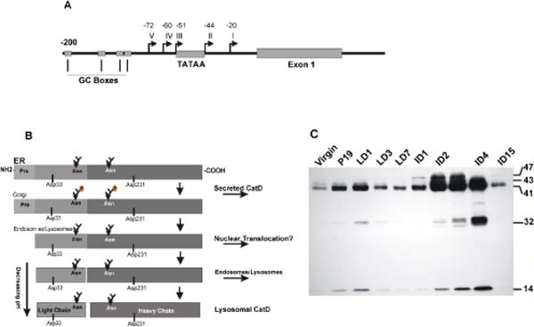
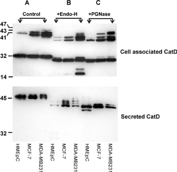
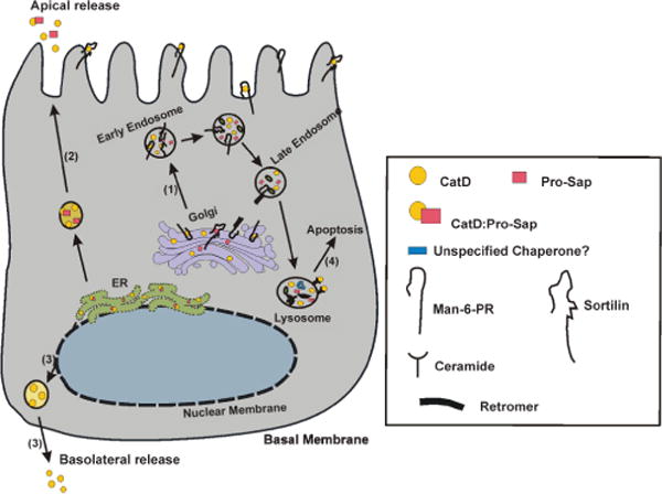
Since its discovery as a lysosomal hydrolase, Cathepsin D (CatD) has been the subject of intensive scrutiny by numerous scientists. Those accumulated efforts have defined its biosynthetic pathway, structure, and companion proteins in the context of its perceived "house keeping" function. However, in the past two decades CatD has emerged as a multifunctional enzyme, involved in myriad biological processes beyond its original "housekeeping" role. CatD is responsible for selective and limited cleavage (quite distinct from non-specific protein degradation) of particular substrates vital to proper cellular function. These proteolytic events are critical in the control of biological processes, including cell cycle progression, differentiation and migration, morphogenesis and tissue remodeling, immunological processes, ovulation, fertilization, neuronal outgrowth, angiogenesis, and apoptosis. Consistent with the biological relevance of CatD, its deficiency, altered regulation or post-translational modification underlie important pathological conditions such as cancer, atherosclerosis, neurological and skin disorders. Specifically, deregulated synthesis, post-translational modifications and hyper-secretion of CatD, along with its mitogenic effects, are established hallmarks of cancer. More importantly, but less studied, is its significance in regulating the sensitivity to anticancer drugs. This review outlines CatD's post-translational modifications, cellular trafficking, secretion and protein binding partners in normal mammary gland, and restates the "site-specific" function of CatD which is most probably dictated by its post-translational modifications and binding partners. Noteworthy, CatD's association with one of its binding partners in the context of drug sensitivity is highlighted, with the optimism that it could contribute to the development of more effective chemotherapeutic agent(s) tailored for individual patients.
Keywords: Binding partners; Cancer; Cathepsin D; Mammary gland; Post-translational modification.
Figures
Similar articles
-
New insights into cathepsin D in mammary tissue development and remodeling.Cancer Biol Ther. 2010 Sep 1;10(5):457-66. doi: 10.4161/cbt.10.5.12534. Epub 2010 Oct 1. Cancer Biol Ther. 2010. PMID: 20592493 Free PMC article.
-
Cleavage of Histone 3 by Cathepsin D in the involuting mammary gland.PLoS One. 2014 Jul 23;9(7):e103230. doi: 10.1371/journal.pone.0103230. eCollection 2014. PLoS One. 2014. PMID: 25054204 Free PMC article.
-
New multienzymatic complex formed between human cathepsin D and snake venom phospholipase A2.J Venom Anim Toxins Incl Trop Dis. 2022 Nov 4;28:e20220002. doi: 10.1590/1678-9199-JVATITD-2022-0002. eCollection 2022. J Venom Anim Toxins Incl Trop Dis. 2022. PMID: 36404954 Free PMC article.
-
Cathepsin D as a Promising Target for the Discovery of Novel Anticancer Agents.Curr Cancer Drug Targets. 2017;17(5):404-422. doi: 10.2174/1568009616666161229145115. Curr Cancer Drug Targets. 2017. PMID: 28215160 Review.
-
Pathophysiological functions of cathepsin D: Targeting its catalytic activity versus its protein binding activity?Biochimie. 2010 Nov;92(11):1635-43. doi: 10.1016/j.biochi.2010.05.009. Epub 2010 May 21. Biochimie. 2010. PMID: 20493920 Review.
Cited by
-
Cathepsins in neuronal plasticity.Neural Regen Res. 2021 Jan;16(1):26-35. doi: 10.4103/1673-5374.286948. Neural Regen Res. 2021. PMID: 32788444 Free PMC article.
-
Autophagy triggers CTSD (cathepsin D) maturation and localization inside cells to promote apoptosis.Autophagy. 2021 May;17(5):1170-1192. doi: 10.1080/15548627.2020.1752497. Epub 2020 Apr 23. Autophagy. 2021. PMID: 32324083 Free PMC article.
-
Lowering Endogenous Cathepsin D Abundance Results in Reactive Oxygen Species Accumulation and Cell Senescence.Mol Cell Proteomics. 2017 Jul;16(7):1217-1232. doi: 10.1074/mcp.M115.050179. Epub 2015 Dec 10. Mol Cell Proteomics. 2017. PMID: 26657266 Free PMC article.
-
Immunotherapy of triple-negative breast cancer with cathepsin D-targeting antibodies.J Immunother Cancer. 2019 Feb 4;7(1):29. doi: 10.1186/s40425-019-0498-z. J Immunother Cancer. 2019. PMID: 30717773 Free PMC article.
-
The Networks of Genes Encoding Palmitoylated Proteins in Axonal and Synaptic Compartments Are Affected in PPT1 Overexpressing Neuronal-Like Cells.Front Mol Neurosci. 2017 Aug 22;10:266. doi: 10.3389/fnmol.2017.00266. eCollection 2017. Front Mol Neurosci. 2017. PMID: 28878621 Free PMC article.
References
-
- Willstätter R, Bamann E. Über die Proteasen der Magenschleimhaut. Erste Abhandlung über die Enzyme der Leukozyten. Hoppe-Seylers Z Physiol Chemie. 1929;180:127–143.
-
- Fruton JS, Irving GW, Bergmann M. On the proteolytic enzymes of animal tissues. II. The composite nature of beef spleen cathepsin. J Biol Chem. 1941;138:249–262.
Grants and funding
LinkOut - more resources
Full Text Sources
Other Literature Sources
Molecular Biology Databases
Miscellaneous