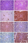
Merkel cell carcinoma in the left groin: A case report and review of the literature
- PMID: 25663881
- PMCID: PMC4315128
- DOI: 10.3892/ol.2015.2861
Merkel cell carcinoma in the left groin: A case report and review of the literature
Abstract
Merkel cell carcinoma (MCC) is a relatively rare but aggressive primary neuroendocrine carcinoma of the skin. MCC frequently occurs on sun-exposed areas in elderly Caucasian patients and has a propensity for local recurrence, regional lymph node invasion and distant metastases. However, MCC occurring on sites that are not sun-exposed in Asian patients is extremely uncommon. The current study describes the case of a 66-year-old Chinese male who presented with an asymptomatic, smooth lesion in the left inguinal region, which was initially diagnosed as a malignant lymphoma. Upon histological and immunohistochemical analysis, the tumor was consistent with the diagnosis of an MCC. In conclusion, due to its low incidence rate and lack of characteristic clinical manifestations, the final diagnosis of MCC relies on the analysis of histological findings and immunohistochemical markers following lesion biopsy or resection. The present study aimed to report a case of MCC and present a brief literature review in order to bring attention to the diagnosis of this condition.
Keywords: Merkel cell carcinoma; differential diagnosis; histopathology; immunohistochemistry; neuroendocrine carcinoma.
Figures

References
-
- Smith DF, Messina JL, Perrott R, et al. Clinical approach to neuroendocrine carcinoma of the skin (Merkel cell carcinoma) Cancer Control. 2000;7:72–83. - PubMed
LinkOut - more resources
Full Text Sources
Other Literature Sources
