. 2015 Mar;22(3):248-55.
doi: 10.1038/nsmb.2964. Epub 2015 Feb 9.

Yersinia effector YopO uses actin as bait to phosphorylate proteins that regulate actin polymerization

Affiliations

Yersinia effector YopO uses actin as bait to phosphorylate proteins that regulate actin polymerization

Wei Lin Lee et al. Nat Struct Mol Biol. 2015 Mar.

Abstract

Pathogenic Yersinia species evade host immune systems through the injection of Yersinia outer proteins (Yops) into phagocytic cells. One Yop, YopO, also known as YpkA, induces actin-filament disruption, impairing phagocytosis. Here we describe the X-ray structure of Yersinia enterocolitica YopO in complex with actin, which reveals that YopO binds to an actin monomer in a manner that blocks polymerization yet allows the bound actin to interact with host actin-regulating proteins. SILAC-MS and biochemical analyses confirm that actin-polymerization regulators such as VASP, EVL, WASP, gelsolin and the formin diaphanous 1 are directly sequestered and phosphorylated by YopO through formation of ternary complexes with actin. This leads to a model in which YopO at the membrane sequesters actin from polymerization while using the bound actin as bait to recruit, phosphorylate and misregulate host actin-regulating proteins to disrupt phagocytosis.


Figures

Figure 1
Figure 1
Structure of the YopO–actin complex. (a) Two views of the structure (front and top view). Actin is shown as a cyan schematic within a gray semitransparent surface with the subdomains labeled. The YopO surface on actin is colored cyan. YopO GDI domain is in red, and the kinase domain is in blue. The catalytic residue Asp267 is shown as magenta spheres. Ordered N- and C-terminal residues are shown as green spheres. Sequence-structural analysis of YopO (89–729) is shown in Supplementary Figure 1. (b) The isolated GDI domain (PDB 2H7O) shown in light orange is superposed on the GDI domain in the YopO–actin complex. At right, YopO is displayed as a surface representation, with the Rac1-binding surface shown in yellow and the actin-binding surface in cyan. The kinase catalytic site (D267) is indicated by an arrow. (c) Domain organization of Y. enterocolitica YopO. (d) Structural elements of the kinase domain of YopO. The αC helix is shown in purple and the catalytic loop in red, with the catalytic Asp267 shown as spheres and the activation segment shown in cyan. Residues involved, or occluded, in the interaction with actin are shown in orange.
Figure 2
Figure 2
The YopO–actin complex is polymerization incompetent. (a) Superimposition of the YopO–actin complex on the actin filament. YopO is colored as in Figure 1a, and the actin filament is represented as a molecular surface. (b) Spectrofluorimetry assay using pyrene-labeled actin to monitor the polymerization of Sf9 actin in the presence of YopO WT and mutants. nRac, non-Rac-binding mutant; nAct, reduced-actin-binding mutant; KD, kinase-dead mutant. (c) Autophosphorylation of YopO WT and mutants in the presence or absence of actin, monitored by autoradiography and Coomassie staining. (d) Pointed-end capping assay of YopO. Tmod, human tropomodulin-3; a.u., arbitrary units; Δ, change.
Figure 3
Figure 3
Structural basis of differential actin isoform preference of YopO. (a) Thr202 of Sf9 actin forms a hydrogen bond with Lys245 on helix α3 of YopO with a bond length of 2.9 Å. YopO is colored as in Figure 1a. (b) Sequence alignment of actins. rACTA, rabbit skeletal α-actin; hACTB, human β-actin; mACTB, mouse β-actin; SfACT, S. frugiperda actin. Thr202 of Sf9 actin is marked with an asterisk. The entire sequence alignment can be found in Supplementary Figure 4. (c) Pyrene-actin polymerization assay for the determination of the monomer sequestration activity of different actin isoforms by YopO WT and K245M. Δ, change; a.u., arbitrary units.
Figure 4
Figure 4
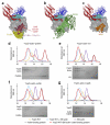
YopO-bound actin interacts with other actin-binding proteins. (a) Model of the quaternary complex of YopO–actin–profilin–VASP fragment, following the color scheme in Figure 1a, with profilin shown in yellow. The surfaces occluded by the actin-binding VASP peptide (red) and profilin-binding polyproline peptide (pink) are colored burgundy and green, respectively. GAB domain, G-actin–binding domain. (b) Model of the tricomplex of YopO–actin–gelsolin, with gelsolin in green. G1–3, gelsolin domains 1–3. (c) Model of the ternary complex of YopO–actin–WH2, with the WH2 peptide of WASP in ocher. Models of the ternary complexes of YopO–actin–G4–6 and YopO–actin–ADFH can be found in Supplementary Figure 5a,b. (dg) Ternary-complex formation of YopO–actin–profilin (d), YopO–actin–G1 (e), YopO–actin–cofilin (f) and YopO–actin–CapG (g), as shown by size-exclusion chromatography. These gel-filtration chromatograms are shown in red and are superimposed on chromatograms of YopO WT, Sf9 actin, YopO WT with Sf9 actin, and the individual actin-binding proteins, colored as indicated. Fractions of the eluted material were analyzed by SDS-PAGE and visualized by Coomassie staining.
Figure 5
Figure 5
Phosphorylation of actin-binding proteins by YopO. (a) In vitro phosphorylation assay, monitored by autoradiography and Coomassie staining. Red asterisks indicate the molecular weights of the substrates. Owing to overlap in the molecular weights of YopO and GST-WASP, the phosphorylated GST-WASP was cleaved with thrombin before SDS-PAGE. Phosphorylation of VASP by various YopO mutants and the quality of the substrates used in the assay can be found in Supplementary Figure 5c,d. (b) Model for phosphorylation of recruited actin-binding proteins by YopO. YopO is colored as in Figure 1a, with actin in gray. The WH2 peptide of WIP (PDB 2A41) is drawn in green, at left. Middle and right show an additional modeled 15-residue peptide binding up and into the kinase active site (colored magenta). (c) Effect of YopO phosphorylation on VASP-mediated actin polymerization. KD, kinase dead.
Figure 6
Figure 6
Model for disabling actin polymerization by YopO. P, phosphorylation; Rac/Rho, Rac and/or Rho.

Comment in

References

    1. Aderem A, Underhill D. Mechanisms of phagocytosis in macrophages. Annu. Rev. Immunol. 1999;17:593–623. - PubMed
    1. Griffin FM, Griffin JA, Leider JE, Silverstein SC. Studies on the mechanism of phagocytosis: requirements for circumferential attachment of particle-bound ligands to specific receptors on the macrophage plasma membrane. J. Exp. Med. 1975;142:1263–1282. - PMC - PubMed
    1. Witke W, Li W, Kwiatkowski DJ, Southwick FS. Comparisons of CapG and gelsolin-null macrophages: demonstration of a unique role for CapG in receptor-mediated ruffling, phagocytosis, and vesicle rocketing. J. Cell Biol. 2001;154:775–784. - PMC - PubMed
    1. Serrander L, et al. Selective inhibition of IgG-mediated phagocytosis in gelsolin-deficient murine neutrophils. J. Immunol. 2000;165:2451–2457. - PubMed
    1. Colucci-Guyon E, et al. A role for mammalian diaphanous-related formins in complement receptor (CR3)-mediated phagocytosis in macrophages. Curr. Biol. 2005;15:2007–2012. - PubMed

Publication types

MeSH terms

Substances

Associated data