

New endoscopic indicator of esophageal achalasia: "pinstripe pattern"
- PMID: 25664812
- PMCID: PMC4321991
- DOI: 10.1371/journal.pone.0101833
New endoscopic indicator of esophageal achalasia: "pinstripe pattern"
Abstract
Background and study aims: Endoscopic diagnosis of esophageal achalasia lacking typical endoscopic features can be extremely difficult. The aim of this study was to identify simple and reliable early indicator of esophageal achalasia.
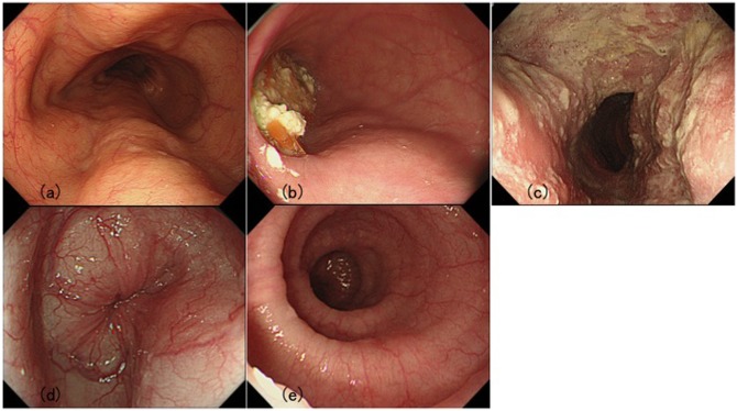
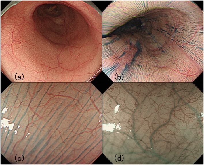
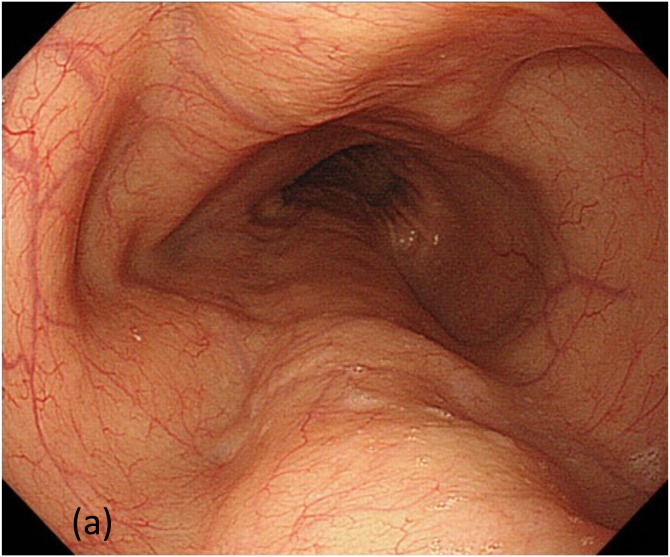
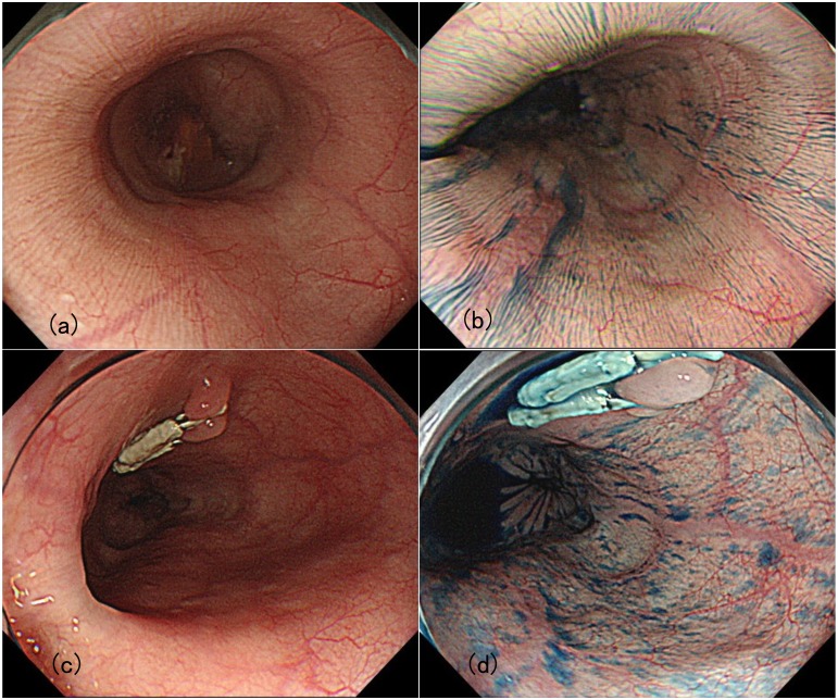
Patients and methods: This single-center retrospective study included 56 cases of esophageal achalasia without previous treatment. As a control, 60 non-achalasia subjects including reflux esophagitis and superficial esophageal cancer were also included in this study. Endoscopic findings were evaluated according to Descriptive Rules for Achalasia of the Esophagus as follows: (1) esophageal dilatation, (2) abnormal retention of liquid and/or food, (3) whitish change of the mucosal surface, (4) functional stenosis of the esophago-gastric junction, and (5) abnormal contraction. Additionally, the presence of the longitudinal superficial wrinkles of esophageal mucosa, "pinstripe pattern (PSP)" was evaluated endoscopically. Then, inter-observer diagnostic agreement was assessed for each finding.
Results: The prevalence rates of the above-mentioned findings (1-5) were 41.1%, 41.1%, 16.1%, 94.6%, and 43.9%, respectively. PSP was observed in 60.7% of achalasia, while none of the control showed positivity for PSP. PSP was observed in 26 (62.5%) of 35 cases with shorter history < 10 years, which usually lacks typical findings such as severe esophageal dilation and tortuosity. Inter-observer agreement level was substantial for food/liquid remnant (k = 0.6861) and PSP (k = 0.6098), and was fair for abnormal contraction and white change. The accuracy, sensitivity, and specificity for achalasia were 83.8%, 64.7%, and 100%, respectively.
Conclusion: "Pinstripe pattern" could be a reliable indicator for early discrimination of primary esophageal achalasia.
Conflict of interest statement
Figures
Similar articles
-
New endoscopic finding of esophageal achalasia with ST Hood short type: Corona appearance.PLoS One. 2018 Jul 31;13(7):e0199955. doi: 10.1371/journal.pone.0199955. eCollection 2018. PLoS One. 2018. PMID: 30063701 Free PMC article.
-
Full-layer mucosal histology in achalasia: Histological epithelial wave is characteristic in "pinstripe pattern"-positive achalasia.Neurogastroenterol Motil. 2018 Jan;30(1). doi: 10.1111/nmo.13168. Epub 2017 Jul 26. Neurogastroenterol Motil. 2018. PMID: 28745833
-
Histology of esophageal mucosa from patients with achalasia.Dis Esophagus. 2005;18(4):257-61. doi: 10.1111/j.1442-2050.2005.00478.x. Dis Esophagus. 2005. PMID: 16128783
-
[Congenital esophageal stenosis owing to ectopic tracheobronchial remnants: report of four cases and review of the literature].Zhonghua Er Ke Za Zhi. 2012 Aug;50(8):571-4. Zhonghua Er Ke Za Zhi. 2012. PMID: 23158732 Review. Chinese.
-
Idiopathic (primary) achalasia: a review.Orphanet J Rare Dis. 2015 Jul 22;10:89. doi: 10.1186/s13023-015-0302-1. Orphanet J Rare Dis. 2015. PMID: 26198208 Free PMC article. Review.
Cited by
-
Usefulness of Endoscopy for the Detection and Diagnosis of Primary Esophageal Motility Disorders and Diseases Relating to Abnormal Esophageal Motility.Diagnostics (Basel). 2023 Feb 12;13(4):695. doi: 10.3390/diagnostics13040695. Diagnostics (Basel). 2023. PMID: 36832183 Free PMC article. Review.
-
Possible new endoscopic finding in patients with achalasia: "Gingko leaf sign".Esophagus. 2020 Apr;17(2):208-213. doi: 10.1007/s10388-019-00684-x. Epub 2019 Jun 21. Esophagus. 2020. PMID: 31227944
-
A clinical study of peroral endoscopic myotomy reveals that impaired lower esophageal sphincter relaxation in achalasia is not only defined by high-resolution manometry.PLoS One. 2018 Apr 2;13(4):e0195423. doi: 10.1371/journal.pone.0195423. eCollection 2018. PLoS One. 2018. PMID: 29608597 Free PMC article.
-
Detection of cytokine storm in patients with achalasia using ELISA.Biomed Rep. 2021 Jul;15(1):62. doi: 10.3892/br.2021.1438. Epub 2021 May 31. Biomed Rep. 2021. PMID: 34113444 Free PMC article.
-
Esophageal Motility Disorders: Current Approach to Diagnostics and Therapeutics.Gastroenterology. 2022 May;162(6):1617-1634. doi: 10.1053/j.gastro.2021.12.289. Epub 2022 Feb 25. Gastroenterology. 2022. PMID: 35227779 Free PMC article. Review.
References
-
- Spiess AE, Kahrilas PJ (1998) Treating achalasia: from whalebone to laparoscope. Jama 280: 638–642. - PubMed
-
- Iwakiri K, Hoshihara Y, Kawami N, Sano H, Tanaka Y, et al. (2010) The appearance of rosette-like esophageal folds ("esophageal rosette") in the lower esophagus after a deep inspiration is a characteristic endoscopic finding of primary achalasia. J Gastroenterol 45: 422–425. 10.1007/s00535-009-0179-7 - DOI - PubMed
-
- Junginger T, Kneist W, Sultanov F, Eckardt VF (2002) [Long-term outcome of myotomy and semi-fundoplication in achalasia]. - PubMed
MeSH terms
LinkOut - more resources
Full Text Sources
Other Literature Sources
Research Materials
Miscellaneous
