

Review
doi: 10.1172/JCI76307.
Epub 2015 Feb 9.
Building a second brain in the bowel
- PMID: 25664848
- PMCID: PMC4362233
- DOI: 10.1172/JCI76307
Item in Clipboard
Review
Building a second brain in the bowel
J Clin Invest.
.
Abstract
The enteric nervous system (ENS) is sometimes called the "second brain" because of the diversity of neuronal cell types and complex, integrated circuits that permit the ENS to autonomously regulate many processes in the bowel. Mechanisms supporting ENS development are intricate, with numerous proteins, small molecules, and nutrients that affect ENS morphogenesis and mature function. Damage to the ENS or developmental defects cause vomiting, abdominal pain, constipation, growth failure, and early death. Here, we review molecular mechanisms and cellular processes that govern ENS development, identify areas in which more investigation is needed, and discuss the clinical implications of new basic research.
Figures
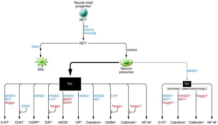
Lineage relationships among enteric neuron subtypes remain poorly understood. This figure summarizes in vivo observations. Gain-of-function data are indicated in red. Loss-of-function data are indicated in blue. Most myenteric neurons arise from TH-negative precursors, as indicated by the relative thickness of arrows.
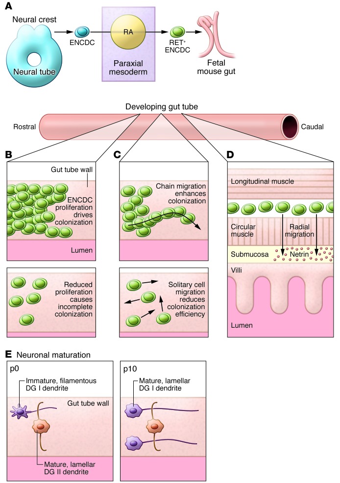
(A) Murine vagal neural crest cells destined for the ENS delaminate from the neural tube at E8.5. These ENCDCs are exposed to RA as they migrate by paraxial mesoderm on their way to the foregut at E9. (B) Once ENCDCs are in developing bowel, efficient caudal migration relies on vigorous ENCDC proliferation (top panel), as disorders that reduce ENCDC proliferation (bottom panel) commonly cause incomplete bowel colonization. (C) Efficient ENCDC migration is facilitated by contact between migrating cells. Chain migration of ENCDCs is quicker and more directed than migration of isolated ENCDCs. Disorders that alter ENCDC cell adhesion also delay bowel colonization and may cause HSCR. (D) After ENCDCs have populated the whole developing bowel (E13.5 in mice) in the region of the future myenteric plexus, a subset of ENCDCs migrates inward radially to form the submucosal plexus. Radial migration is regulated by the RET-GDNF signaling axis and by netrin/DCC chemoattraction. (E) nNOS-IR DG I neurons send caudal projections in the longitudinal axis, whereas CGRP-IR DG II neurons project circumferentially. Both DG I and DG II neurons are present at P0. However, only DGII neurons exhibit mature lamellar dendrites at this age, whereas most DG I dendrites are still filamentous. The proportion of DG I lamellar dendrites increases from P0 to P10. DG II projections do not grow in length from P0 to P10, whereas DG I projections do, though their growth rate does not match that of the bowel. There is significant maturation of the ENS after birth, at least in rodents.
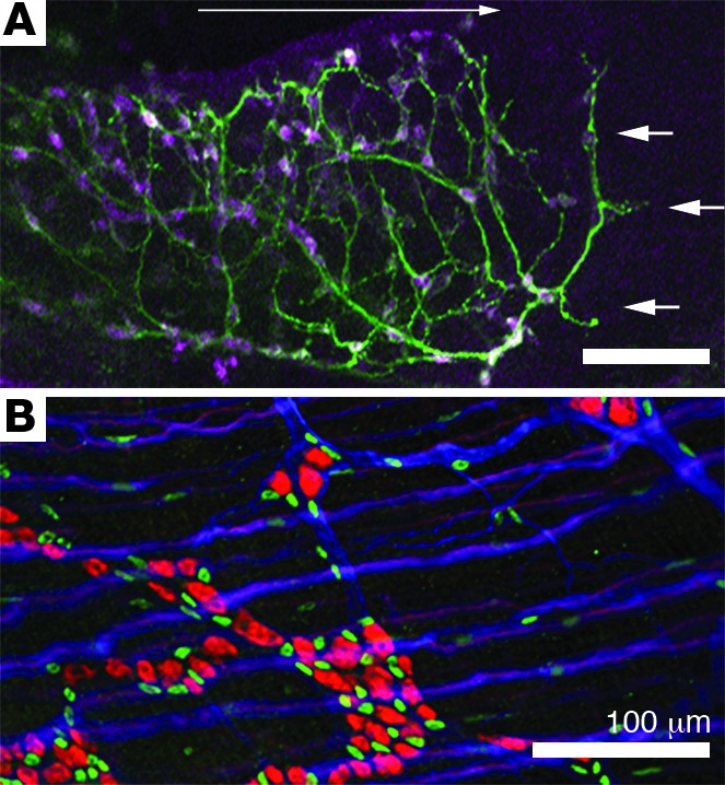
(A) Vagal ENCDCs migrate in a rostral to caudal direction through fetal bowel (long white arrow). At E12.5 ENS precursors have migrated halfway through the fetal colon. ANNA-1 antibody binds HuC/D antigen and identifies enteric neurons (magenta), while TuJ1 binds neuron-specific β-III tubulin and labels neurites (green). ENCDCs migrate in chains though the bowel, but during the period of migration some precursors differentiate into neurons and extend neurites, including at the migration wavefront (white arrows). (B) Adult small bowel myenteric plexus, indicated by ANNA-1 antibody (red, neurons), SOX10 antibody (green, enteric glia), and TuJ1 antibody (blue), demonstrates clusters of neurons and glia in mature ganglia as well as many neurites within and between ganglia. Scale bars: 100 microns.
References
-
- Wood JD. Taming the irritable bowel. Curr Pharm Des. 2013;19(1):142–156. - PubMed
-
- Heuckeroth RO. Hirschsprung disease. In: Faure C, DiLorenzo C, Thapar N, eds. Pediatric Neurogastroenterology: Gastrointestinal Motility And Functional Disorders In Children. New York, New York, USA: Springer; 2013:271–283.
