. 2015 Mar 2;125(3):1215-27.
doi: 10.1172/JCI76693. Epub 2015 Feb 9.

Annexin A1-containing extracellular vesicles and polymeric nanoparticles promote epithelial wound repair

Annexin A1-containing extracellular vesicles and polymeric nanoparticles promote epithelial wound repair

Giovanna Leoni et al. J Clin Invest. .

Abstract

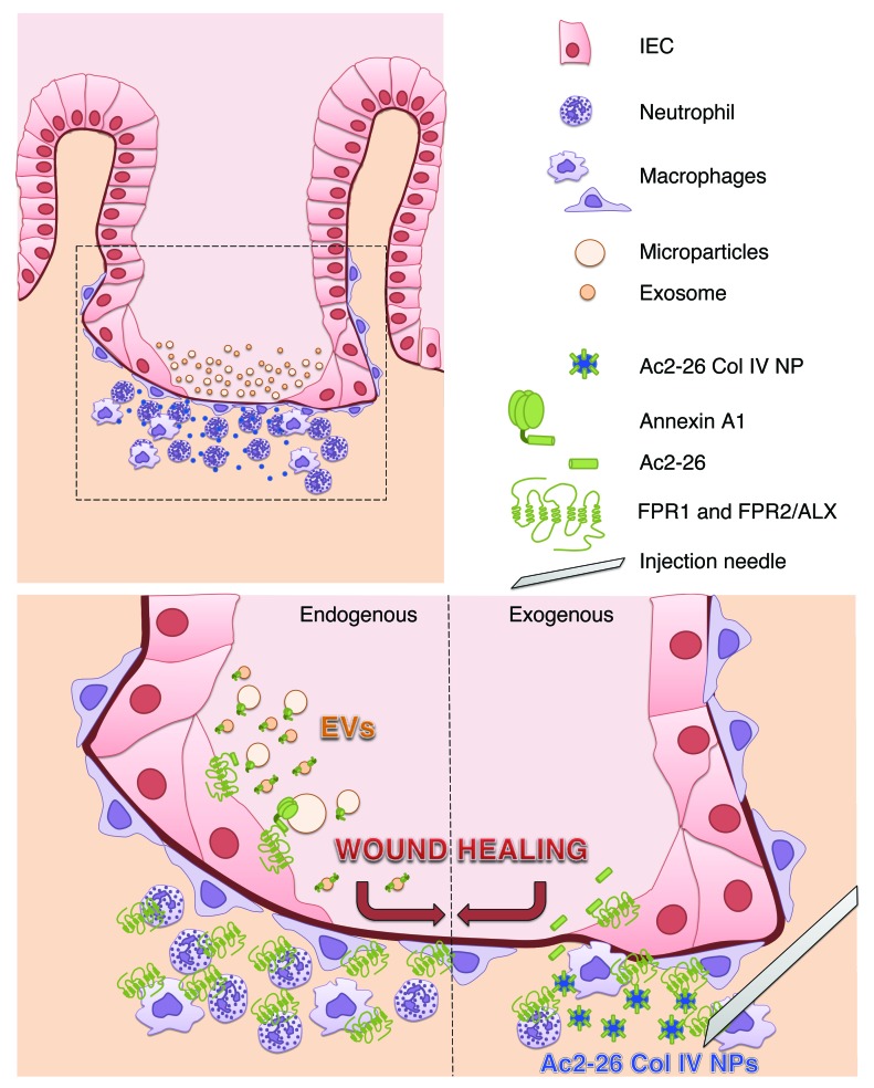
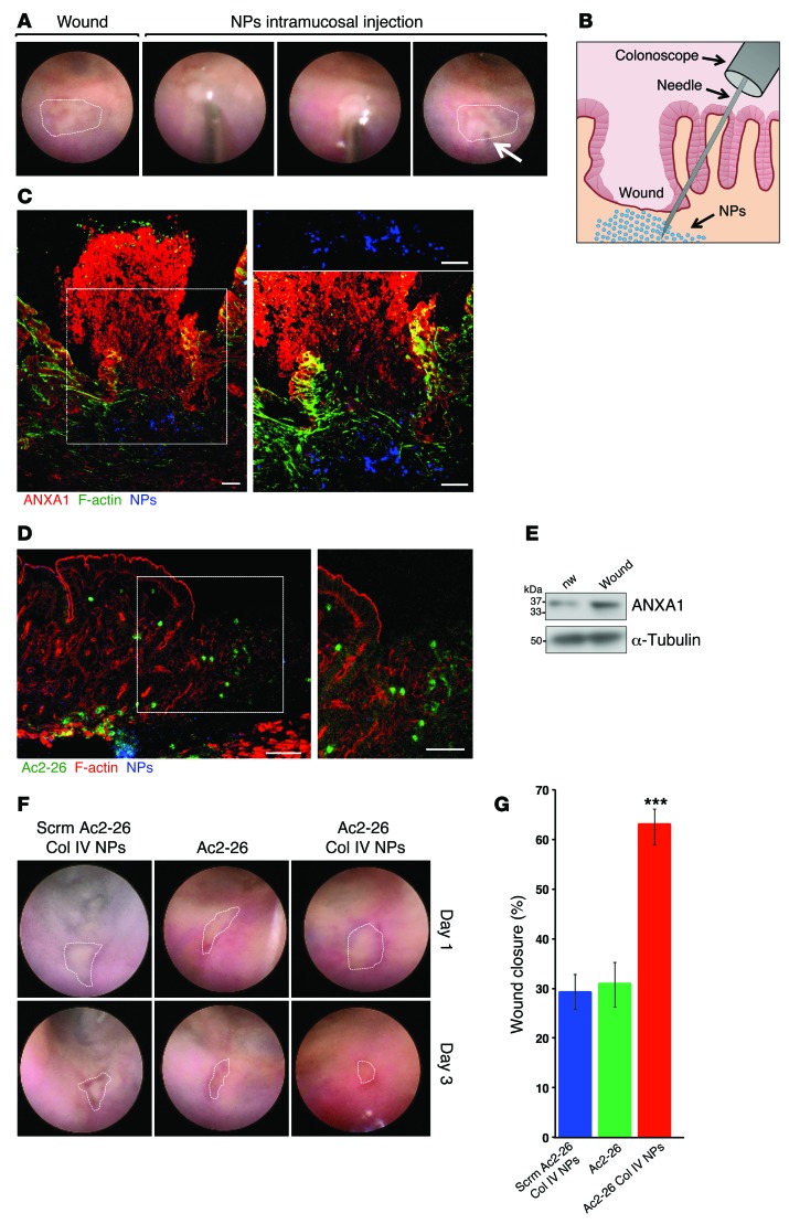
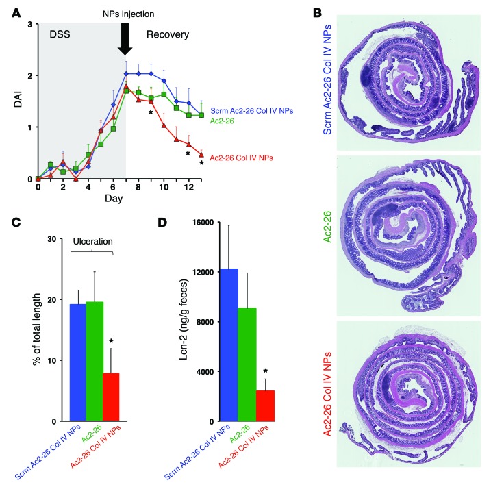
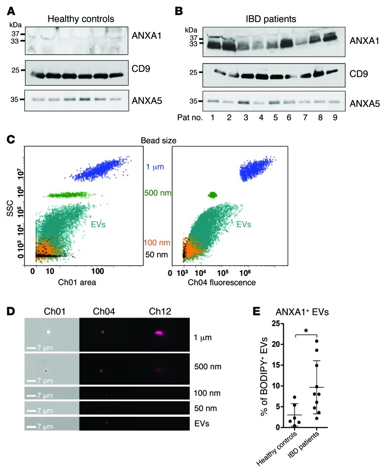
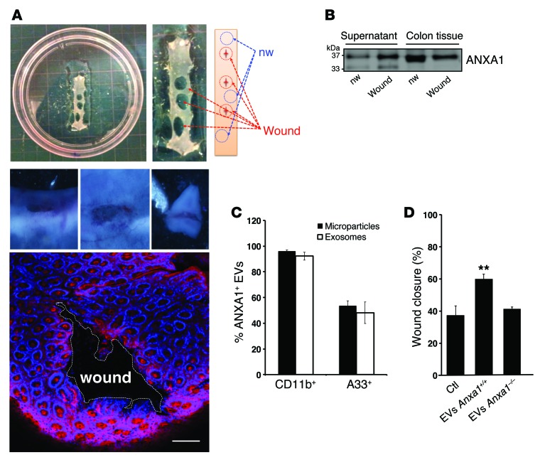
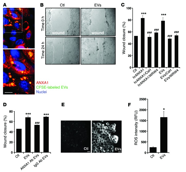
Epithelial restitution is an essential process that is required to repair barrier function at mucosal surfaces following injury. Prolonged breaches in epithelial barrier function result in inflammation and further damage; therefore, a better understanding of the epithelial restitution process has potential for improving the development of therapeutics. In this work, we demonstrate that endogenous annexin A1 (ANXA1) is released as a component of extracellular vesicles (EVs) derived from intestinal epithelial cells, and these ANXA1-containing EVs activate wound repair circuits. Compared with healthy controls, patients with active inflammatory bowel disease had elevated levels of secreted ANXA1-containing EVs in sera, indicating that ANXA1-containing EVs are systemically distributed in response to the inflammatory process and could potentially serve as a biomarker of intestinal mucosal inflammation. Local intestinal delivery of an exogenous ANXA1 mimetic peptide (Ac2-26) encapsulated within targeted polymeric nanoparticles (Ac2-26 Col IV NPs) accelerated healing of murine colonic wounds after biopsy-induced injury. Moreover, one-time systemic administration of Ac2-26 Col IV NPs accelerated recovery following experimentally induced colitis. Together, our results suggest that local delivery of proresolving peptides encapsulated within nanoparticles may represent a potential therapeutic strategy for clinical situations characterized by chronic mucosal injury, such as is seen in patients with IBD.


Figures

Figure 7
Figure 7. Endogenous epithelial-derived ANXA1-containing EVs and exogenous administration of Ac2-26 Col IV NPs activate FPRs to promote wound repair.
Epithelial cells adjoining the wound flatten out, change polarity, and migrate as a sheet to cover denuded surfaces. Immune cells infiltrate into the site of injury in mucosal wounds. The healing epithelium is exposed to infiltrating immune cells that coordinate the mucosal repair. During this process, epithelial cells and immune cells release EVs (microparticles and exosomes) containing proresolution mediators, including ANXA1. ANXA1 in EVs signals through epithelial FPRs (FPR1 and FPR2/ALX) to promote wound repair (acting as an endogenous mediator of wound repair). This prorepair effect of endogenous ANXA1 can be mimicked using synthetically derived NPs containing ANXA1 mimetic peptide Ac2-26. Intramucosal injection of Ac2-26 Col IV NPs enhances epithelial wound healing (after exogenous administration of ANXA1).
Figure 6
Figure 6. Intramucosal injections of Ac2-26 Col IV NPs augment colonic mucosal wound healing.
(A) Images obtained during colonoscopy showing intramucosal injection of NPs in the mucosal wound bed. The arrow delineates the area of submucosal injection. (B) Schematic showing intramucosal NP injection into the colonic mucosal wounds. (C) Frozen sections of resealing colonic wounds in Anxa1+/+ mice showing ANXA1 (red), F-actin (Alexa Fluor 488 phalloidin, green), and NPs (Alexa Fluor 647, blue). Higher-magnification images (original magnification, ×40) of the boxed region (original magnification, ×20) are shown to the right. Scale bar: 50 μm. (D) Frozen sections labeled for F-actin (Alexa Fluor 555 phalloidin, red). Released Ac2-26-FAM is shown in green and labeled Ac2-26 Col IV NPs are shown in blue. A higher-magnification image of the boxed region is shown to the right. Scale bar: 50 μm. (E) Immunoblot analysis of ANXA1 expression in nonwounded and resealing wounds of Anxa1+/+ mice. Equivalent amounts of protein were loaded. From densitometry analysis ANXA1 protein in wounded tissue is increased versus nonwounded cells (2.568-fold increase, P = 0.0005, average of n = 3 immunoblots). (F) Endoscopic images of healing colonic mucosal wounds 1 or 3 days after biopsy-induced injury in Anxa1–/– mice treated with NPs. (G) Quantification of wound repair. Data are expressed as mean ± SEM; n = 15 mice per group. ***P < 0.0001 vs. Scrm Ac2-26 Col IV NPs and vs. Ac2-26. Statistical comparisons were performed by ANOVA with Tukey’s multiple comparison post-test.
Figure 5
Figure 5. NPs containing Ac2-26 accelerate recovery of murine colitis in vivo.
(A) Clinical disease activity index (DAI) of Anxa1–/– mice subjected to DSS colitis (7 days) followed by recovery from colitis (6 days). NPs and Ac2-26 peptide alone were administered only once via intraperitoneal injection (4 μg per mouse) at the first day of recovery (*P < 0.001). (B) Representative photomicrographs of hematoxylin and eosin–stained histological sections (original magnification, ×2). (C) Analysis of percentage of ulceration in whole colon samples (*P = 0.0131 vs. Scrm Ac2-26 Col IV NPs). (D) Fecal lipocalin analysis (*P = 0.0037 vs. Scrm Ac2-26 Col IV NPs; n = 10 mice per group). Quantitative data are expressed as mean ± SEM for each treatment group. Statistical comparisons were performed by ANOVA with Tukey’s multiple comparison post-test.
Figure 4
Figure 4. Serum-derived exosomes from healthy controls and patients with IBD.
(A and B) Immunoblots of ANXA1, CD9, and ANXA5 in isolated exosomes from sera of human (A) healthy controls and (B) patients with IBD. Exosomes were precipitated from human pooled sera using SBI’s Serum ExoQuick precipitation reagent (data are representative of n = 3 experiments). (C) Representative side scatter versus bright-field area (SSC/Ch12) and Ch04 fluorescence plots for EV isolates acquired with 1-μm and 500-nm reference beads (100-nm and 50-nm beads were acquired separately and overlaid) of EVs isolated from human sera by ultracentrifugation (n = 3). (D) Representative bright-field (Ch01), Ch04, and Ch012 images of each population of beads and EVs (n = 3) (original magnification, ×60). Scale bar: 7 μm. (E) EVs were stained with BODIPY-Texas Red and conjugated fluorescent antibodies against ANXA1 (n = 3). Data are expressed as mean ± SD. *P = 0.0194. Statistical comparisons were performed by 2-tailed Student’s t test.
Figure 3
Figure 3. Analysis of ANXA1-containing EVs in ex vivo cultures of resealing mucosal wounds and functional effects of EVs on wound closure.
(A) 4-mm punch biopsy of resealing colonic wounds (1 day after injury, red circle) and nonwounded mucosa (blue circle) were rinsed in sterile PBS to remove cellular debris and immediately placed into a 24-well culture plate (3 wounds from each mouse per well, n = 4). Photographic images of a wound (1- to 2-mm size) and confocal images of resealing mucosal wound showing F-actin (Alexa Fluor 555 phalloidin, pink) and nuclei (TO-PRO-3, blue) are shown (n = 4). Scale bar: 100 μm. (B) Immunoblot showing ANXA1 in the supernatant and lysate of the ex vivo culture. Densitometry analysis shows that ANXA1 protein in culture supernatants derived from wounded cultures is significantly increased versus nonwounded cells (1.979-fold increase, P = 0.0024 average of n = 3 immunoblots). In total lysate, the ratio of ANXA1 expression in wounded tissue versus nonwounded tissue is 0.762 (P = 0.0005 average of n = 3 immunoblots). (C) EVs (microparticles and exosomes) were stained with BODIPY-Texas Red and conjugated CD11b, A33, and ANXA1 fluorescent antibodies. Data are expressed as mean ± SEM; n = 4 per group. (D) EVs derived from Anxa1+/+ and Anxa1–/– mice were added to monolayers of cultured murine epithelial cells, and scratch wound resealing assay was performed. Increased wound closure was observed after incubation with ANXA1-containing EVs (**P = 0.0025) compared with EVs lacking ANXA1. Mean ± SEM; n = 15 mice per group. Statistical comparisons were performed by ANOVA with Tukey’s multiple comparison post-test.
Figure 2
Figure 2. EVs containing ANXA1 regulate epithelial wound healing.
(A) Representative (of n = 5) en face image of migrating IECs after 24 hours of interaction with labeled EVs (CFSE, 10 μmol/l; Molecular Probes). The boxed area (original magnification, ×40) is shown at high magnification in the image below (original magnification, ×100). Scale bar: 10 μm. (B and C) Scratch wound healing assay using IEC monolayers. EVs and hrANXA1 were added to wounded IECs alone and in the presence of FPR1 antagonist, CsH (1 μM); FPR2/ALX antagonist, WRW4 (10 μM); and other conditions, as indicated. Wound widths were determined at time 0 and 24 hours (n = 5, mean ± SEM) (original magnification, ×10). (D) Scratch wound healing assay using IEC monolayers. EVs were added to wounded IECs alone and in the presence of mouse monoclonal ANXA1 inhibitory antibody (250 μg/ml) or control IgG antibody (250 μg/ml). The experiments in C and D were repeated 3 times, and results of one representative experiment performed with 5 parallel samples are shown (mean ± SEM). ***P < 0.0001 compared with control; ###P < 0.0001 compared with EVs. (E and F) IECs were incubated with EVs for 15 minutes, and ROS generation was detected by confocal microscopy using the fluorescent Hydro-Cy3 dye (15 μM). Scale bar: 40 μm. Summarized data for Hydro-Cy3 fluorescence intensity are presented in the graph (mean ± SEM, *P = 0.0002 vs. control, n = 3). Confocal micrographs are representative of 3 independent experiments. All the results in this figure are representative of at least 3 independent experiments. Statistical comparisons were performed by ANOVA with Tukey’s multiple comparison post-test for C and D and 2-tailed Student’s t test for F. RFU, relative fluorescent units.
Figure 1
Figure 1. Intestinal epithelial wounding induces release of ANXA1-bearing EVs.
(A) Immunoblotting for ANXA1 protein in supernatant of nonwounded (nw) or wounded (wound) human epithelial cells. Densitometry analysis shows that ANXA1 protein in cell supernatants derived from wounded cells is significantly increased versus nonwounded confluent cells (2.94-fold increase, P < 0.0001, average of n = 5 immunoblots), and no significant change was observed in the lysate. (B) Immunoblotting for ANXA1 protein in EVs isolated from supernatant. Densitometry analysis shows that ANXA1 protein on EVs derived from wounded cells is increased versus nonwounded cells (2.166-fold increase, P = 0.0066, average of n = 4 immunoblots). (C) Immunogold labeling for ANXA1 and transmission electron microscopy to detect ANXA1 distribution in EVs. Scale bar: 100 nm. (D) Immunoblotting for conventional EV markers in epithelial cell lysates (representative of n = 4 immunoblots). sADAM-10, soluble ADAM-10. (E) EVs were incubated with CD63+ Dynabeads and analyzed by immunoblotting. Densitometry analysis shows that ANXA1 protein on EVs derived from wounded cells is increased versus nonwounded cells (1.459-fold increase, P = 0.0004, average of n = 4 immunoblots). (F) Biotinylated cell surface proteins released from the supernatant of nonwounded and wounded IECs captured with avidin sepharose beads. Densitometry analysis shows that ANXA1 protein on EVs derived from wounded cells is increased versus nonwounded cells (4.239-fold increase increase, P = 0.0002, average of n = 4 immunoblots). (G) Immunoblots show ANXA1 and CD9 proteins in EVs after IEC wounding and treatment with BAPTA-AM (30 μM), Z-VAD-FMK (25 mM), cytochalasin D (2 μM), jasplakinolide (1 μM), dynasore (80 μM), ML-7 (20 μM), and Y-27632 (10 μM). All the results in this figure are representative of at least 3 independent experiments.

References

    1. Koch S, Nusrat A. The life and death of epithelia during inflammation: lessons learned from the gut. Annu Rev Pathol. 2012;7:35–60. doi: 10.1146/annurev-pathol-011811-120905. - DOI - PubMed
    1. Serhan CN, Dalli J, Colas RA, Winkler JW, Chiang N. Protectins and maresins: new pro-resolving families of mediators in acute inflammation and resolution bioactive metabolome. Biochim Biophys Acta. doi: 10.1016/j.bbalip.2014.08.006. [published online ahead of print August 17, 2014]. doi: 10.1016/j.bbalip.2014.08.006. - DOI - PMC - PubMed
    1. Serhan CN. Pro-resolving lipid mediators are leads for resolution physiology. Nature. 2014;510(7503):92–101. doi: 10.1038/nature13479. - DOI - PMC - PubMed
    1. Babbin BA, et al. Annexin I regulates SKCO-15 cell invasion by signaling through formyl peptide receptors. J Biol Chem. 2006;281(28):19588–19599. doi: 10.1074/jbc.M513025200. - DOI - PubMed
    1. Martin GR, Perretti M, Flower RJ, Wallace JL. Annexin-1 modulates repair of gastric mucosal injury. Am J Physiol Gastrointest Liver Physiol. 2008;294(3):G764–G769. doi: 10.1152/ajpgi.00531.2007. - DOI - PubMed

Publication types