. 2015 Apr;35(8):1414-32.
doi: 10.1128/MCB.01501-14. Epub 2015 Feb 9.

Role of the unfolded protein response in regulating the mucin-dependent filamentous-growth mitogen-activated protein kinase pathway

Affiliations

Role of the unfolded protein response in regulating the mucin-dependent filamentous-growth mitogen-activated protein kinase pathway

Hema Adhikari et al. Mol Cell Biol. 2015 Apr.

Abstract

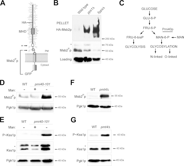
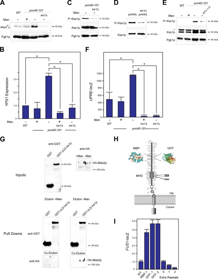
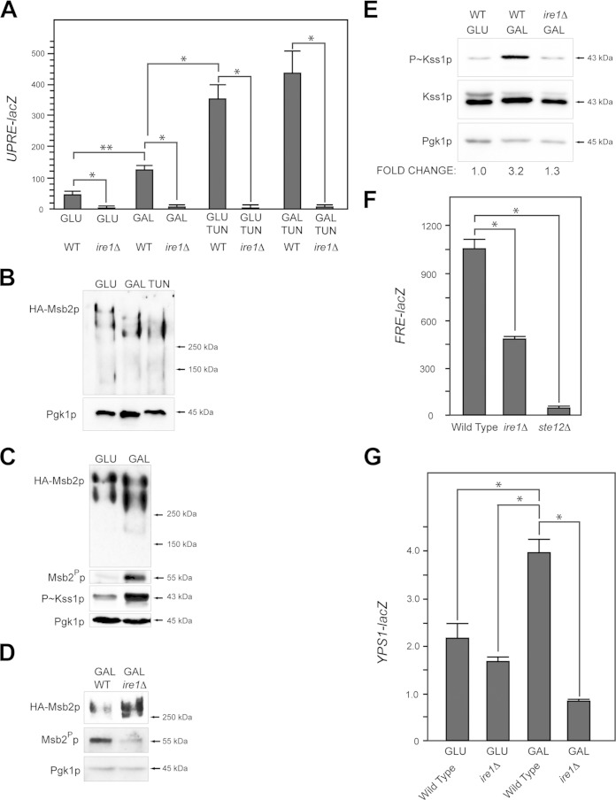
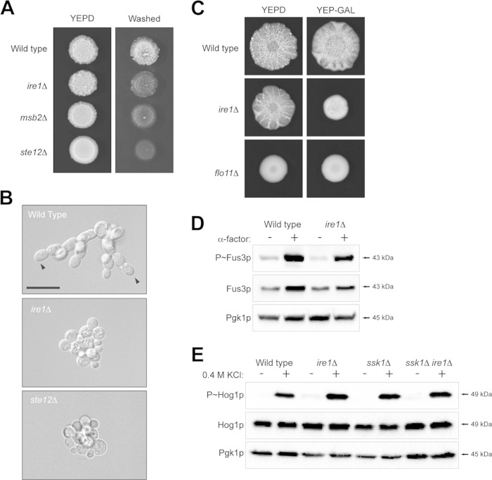
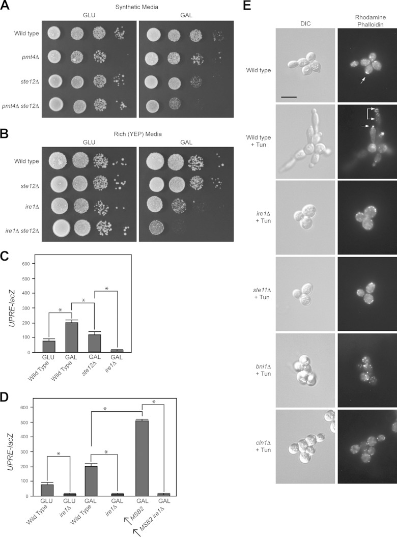
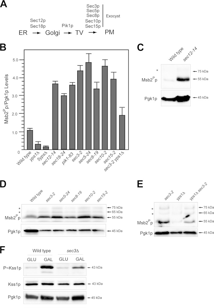
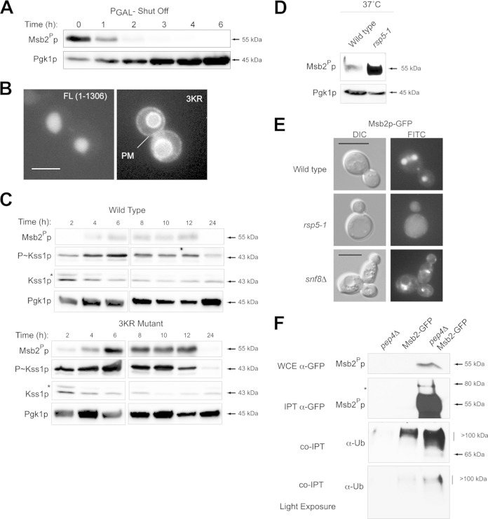
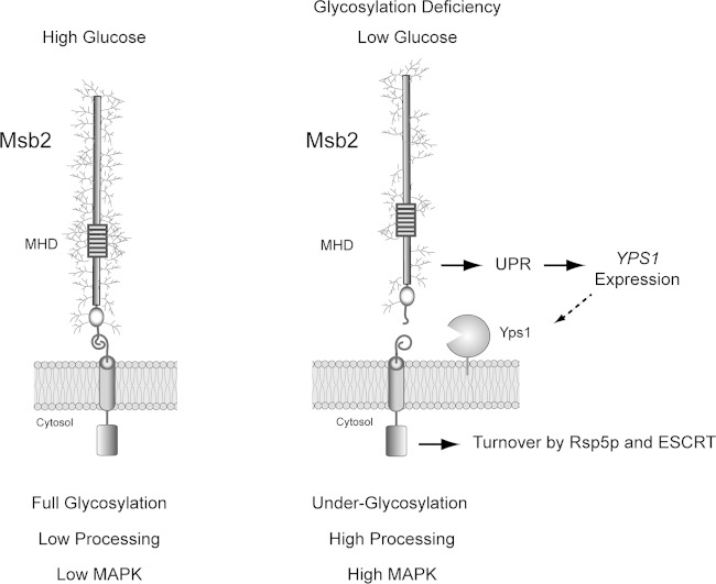
Signaling mucins are evolutionarily conserved regulators of signal transduction pathways. The signaling mucin Msb2p regulates the Cdc42p-dependent mitogen-activated protein kinase (MAPK) pathway that controls filamentous growth in yeast. The cleavage and release of the glycosylated inhibitory domain of Msb2p is required for MAPK activation. We show here that proteolytic processing of Msb2p was induced by underglycosylation of its extracellular domain. Cleavage of underglycosylated Msb2p required the unfolded protein response (UPR), a quality control (QC) pathway that operates in the endoplasmic reticulum (ER). The UPR regulator Ire1p, which detects misfolded/underglycosylated proteins in the ER, controlled Msb2p cleavage by regulating transcriptional induction of Yps1p, the major protease that processes Msb2p. Accordingly, the UPR was required for differentiation to the filamentous cell type. Cleavage of Msb2p occurred in conditional trafficking mutants that trap secretory cargo in the endomembrane system. Processed Msb2p was delivered to the plasma membrane, and its turnover by the ubiquitin ligase Rsp5p and ESCRT attenuated the filamentous-growth pathway. We speculate that the QC pathways broadly regulate signaling glycoproteins and their cognate pathways by recognizing altered glycosylation patterns that can occur in response to extrinsic cues.


Figures

FIG 1
FIG 1
Underglycosylated Msb2p is proteolytically processed at elevated levels. (A) The Msb2p protein is shown as a single-pass glycoprotein with the mucin homology domain (MHD). Cleavage sites corresponding to immunoblot data for cleaved Msb2p-GFP are indicated by arrows. Msb2Pp refers to the proteolytically processed form; the asterisk refers to a minor cleavage product. The positions of HA and GFP fusions are shown. (B) Cleavage of HA-Msb2p-GFP in the yps1Δ and 5ypsΔ mutants. The top blot was probed with anti-HA antibodies to show full-length Msb2p at >250 kDa (HA-Msb2p). The middle blot was probed with anti-GFP antibodies to show the proteolytically processed Msb2Pp-GFP fusion (Msb2Pp, 55 kDa; *, 75 kDa). Blots were probed with anti-Pgk1p antibody, which was used as a loading control for all experiments. (C) Pathway for the conversion of glucose into substrates for glycolysis and protein glycosylation. The Pmi40p enzyme is underlined. (D) Immunoblot of Msb2Pp in wild-type cells (WT) and the pmi40-101 mutant grown in YEPD (−Man) or YEPD plus 50 mM mannose (+Man) for 5.5 h. (E) Immunoblot of P∼Kss1p levels for the strains used in panel D. The asterisk refers to a background band seen under some conditions with the total Kss1p antibodies. (F) Msb2Pp levels in wild-type (WT) cells and the pmt4Δ mutant. Cells were grown in YEP-GAL for 6 h. (G) P∼Kss1p levels for the strains examined in panel F.
FIG 2
FIG 2
Ire1p regulates Msb2p cleavage and filamentous-growth pathway activity by regulating YPS1 expression in response to protein glycosylation deficiency. (A) Msb2p cleavage in wild-type cells and the pmi40-101 mutant grown with (YEPD + Man) or without (YEPD) mannose in combination with the ire1Δ mutant. (B) YPS1 expression was determined by qPCR and adjusted to ACT1 levels as a control. The indicated strains were grown in YEPD (−Man) or YEPD plus Man medium (+Man) for RNA preparation and qPCR analysis. The asterisk refers to a P value of <0.05. (C) P∼Kss1p levels in the pmi40-101 and pmi40-101 ire1Δ double mutant. (D) P∼Kss1p levels in the pmt4Δ and pmt4Δ ire1Δ double mutants grown in YEP-GAL. (E) P∼Kss1p levels for an Ire1p C-terminal truncation. (F) UPRE-lacZ activity was determined by β-galactosidase assays for the indicated strains and conditions. Experiments were performed in duplicate, and the average values are shown. Error bars represent the standard deviations between trials. The asterisk refers to a P value of <0.05. (G) In vitro pulldown of HA-Msb2p expressed in the pmi40-101 mutant in YEPD (with or without mannose) with the luminal domain of Ire1p, called cLD-Ire1p-GST. Input, pulldown, and coelutions are shown. (H) Msb2p with insertion of tandem repeats, MBP, or GFP shown. GFP-1 was inserted at residue 324, resulting in an in-frame deletion of aa 324 to 326. GFP-2 was inserted at residue 246 and resulted in a deletion of aa 246 to 539. MPB was inserted at residue 324 without deletion of amino acid residues. (I) MAPK activity was assessed by an Msb2p-dependent reporter (FUS1-lacZ, which in Σ1278b ste4 strains shows Msb2p dependence [15]). Differences are expressed as fold differences compared to those of wild-type cells. Error bars represent standard deviations between trials, which varied by less than 10%.
FIG 3
FIG 3
Ire1p regulates cleavage of Msb2p and the filamentous-growth pathway during growth in galactose. (A) UPRE-lacZ activity for the indicated strains and conditions (TUN, 2.5 μg/ml tunicamycin). The experiment was performed in duplicate. Error bars represent the standard deviations between trials. *, P < 0.05; **, P < 0.09. (B) Immunoblot of elutions from ConA columns showing HA-Msb2p migration on low-percentage acrylamide gels (6% SDS-PAGE) under the indicated conditions. (C) Immunoblots of HA-Msb2p migration (top, anti-HA, 6% SDS-PAGE gel), Msb2p cleavage (anti-GFP immunoblot Msb2Pp), P∼Kss1p levels, and total protein levels (anti-Pgk1p) of extracts from cells grown in glucose (GLU, YEPD), galactose (GAL, YEP-GAL), or TUN (YEPD plus 2.5 μg/ml tunicamycin). (D) Immunoblots showing Msb2p cleavage in wild-type cells (WT) and the ire1Δ mutant. The anti-HA antibodies were used to evaluate HA-Msb2p, and GFP antibodies were used to evaluate Msb2Pp-GFP. (E) P∼Kss1p levels in wild-type cells (WT) and the ire1Δ mutant incubated in YEPD (GLU) and YEP-GAL (GAL) medium. Numbers refer to fold differences relative to the loading control determined by assessing band intensity by ImageJ. (F) FRE-lacZ expression in the wild type and ire1Δ and ste12Δ mutants grown in YEP-GAL for 12 h. The experiment was performed in duplicate. Error bars represent the standard deviations between experiments. *, P < 0.05. (G) YPS1-lacZ expression in the wild-type and ire1Δ strains in YEPD (GLU) and YEP-GAL (GAL) medium at 24 h. The experiment was performed in duplicate. Error bars represent the standard deviations between experiments. *, P < 0.05.
FIG 4
FIG 4
Role of Ire1p in regulating invasive growth, biofilm/mat formation, and other MAPK pathways that share components with the filamentous-growth pathway. (A) Wild-type, msb2Δ, ire1Δ, and ste12Δ strains were spotted onto YEPD medium. After 48 h, the plate was photographed, washed, and photographed again to reveal invaded cells. (B) Single-cell assay showing the growth after 16 h of strains on synthetic medium lacking glucose. Arrowheads refer to examples of distal-unipolar buds. Bar, 15 μm. (C) Biofilm/mat formation. Wild-type cells and ire1Δ and flo11Δ mutant cells were spotted on YEPD and YEP-GAL media (0.3% agar) for 3 days. (D) Phosphorylation of Fus3p in response to α-factor in wild-type cells and the ire1Δ mutant. (E) Phosphorylation of Hog1p in wild-type cells and the ire1Δ mutant exposed to 0.4 M KCl for 10 min in wild-type cells and an ssk1Δ background.
FIG 5
FIG 5
Roles of UPR in regulating polarity and growth in poor carbon sources. (A) Serial dilutions were spotted onto synthetic medium containing glucose or galactose. The ste12Δ mutant showed a modest growth defect on synthetic media with galactose. (B) Serial dilutions were spotted onto YEPD and YEP-GAL media. (C) UPRE-lacZ activity of the indicated strains in YEPD (GLU) and YEP-GAL (GAL). The experiment was performed in duplicate; error bars show standard deviations between strains. *, P < 0.05. (D) UPRE-lacZ activity was performed as described for panel C. The arrow refers to Msb2p overexpressed by the pGAL1 promoter. (E) Rhodamine-phalloidin staining of wild-type cells and the indicated mutants grown to saturation in YEPD medium. Tun, tunicamycin. Bar, 5 μm.
FIG 6
FIG 6
Proteolytic processing of Msb2p in protein-trafficking mutants. (A) Proteins that regulate trafficking in the secretory pathway. Mutants were examined at the nonpermissive temperature (4 h of growth in YEPD at 37°C) to arrest protein trafficking at different points along the secretory pathway. TV, transit vesicles. (B) Quantitation of Msb2Pp adjusted to total protein levels for the indicated mutants. The Msb2Pp/Pgk1p ratio for wild-type cells was set to 1 and compared to those of other mutants, assessed by band intensity by immunoblot analysis and analyzed by ImageJ. Intensities varied less than 10% between trials. (C) Msb2p cleavage in the sec12-14 mutant. Asterisks refer to minor cleavage products for panels C to E. (D) Msb2p cleavage in exocyst mutants. (E) Msb2p cleavage in sec3-2 yps1Δ mutant alongside control strains. (F) P∼Kss1p levels in wild-type cells and the sec3Δ mutant incubated for 4 h in YEPD (GLU) or YEP-GAL (GAL).
FIG 7
FIG 7
Roles for Rsp5p and ESCRT in regulating the turnover of Msb2p. (A) Promoter shutoff showing Msb2Pp levels at the indicated time points. (B) Localization of full length (FL; aa 1 to 1306) and Msb23KRp-GFP (3KR). Bar, 5 μm. (C) Role of lysines in the turnover domain of Msb2p in impacting the stability of the protein and MAPK activity. (Top) Msb2p-GFP and Msb23KRp-GFP levels over a culture-growth cycle at the indicated time points. (Middle) P∼Kss1p activity. (Bottom) Pgk1p levels. Proteins also were examined side by side on separate blots to directly compare protein levels. (D) Msb2Pp-GFP levels in wild-type cells and in mutants harboring temperature-sensitive rsp5 alleles. Strains were grown at the nonpermissive temperature (37°C) and evaluated by immunoblot analysis. (E) The localization of Msb2p-GFP in wild-type cells, the rsp5-1 mutant (at 37°C), and the snf8Δ (ESCRT) mutant. (F) Msb2p-GFP was immunoprecipitated (IPT) from wild-type cells and the pep4Δ mutant, and extracts were probed using anti-GFP and anti-UB antibodies. Stabilization of Msb2p in the pep4Δ mutant resulted in higher levels of the ubiquitin-modified forms of the protein. Bottom panel, lighter exposure. Ub, ubiquitin; WCE, whole-cell extract.
FIG 8
FIG 8
Role of the UPR in regulating Msb2p cleavage and activation of the filamentous-growth pathway. (Left) During growth in glucose-replete conditions, Msb2p is fully glycosylated. As a result, Msb2p is not efficiently processed and MAPK activity is low. (Right) During growth in poor carbon sources (like galactose) or in cells experiencing a protein glycosylation deficiency, Msb2p becomes underglycosylated. Underglycosylated Msb2p is recognized by Ire1p, a major regulator of the UPR. Ire1p regulates expression of YPS1 (dashed arrow), resulting in elevated cleavage of Msb2p and activation of the filamentous-growth pathway. The proteolytically processed form of Msb2p is turned over by Rsp5p and ESCRT to attenuate the filamentous-growth pathway.

References

    1. Kufe DW. 2009. Mucins in cancer: function, prognosis and therapy. Nat Rev Cancer 9:874–885. doi: 10.1038/nrc2761. - DOI - PMC - PubMed
    1. Tian E, Ten Hagen KG. 2009. Recent insights into the biological roles of mucin-type O-glycosylation. Glycoconj J 26:325–334. doi: 10.1007/s10719-008-9162-4. - DOI - PMC - PubMed
    1. Bafna S, Kaur S, Batra SK. 2010. Membrane-bound mucins: the mechanistic basis for alterations in the growth and survival of cancer cells. Oncogene 29:2893–2904. doi: 10.1038/onc.2010.87. - DOI - PMC - PubMed
    1. Cullen PJ. 2011. Post-translational regulation of signaling mucins. Curr Opin Struct Biol 21:590–596. doi: 10.1016/j.sbi.2011.08.007. - DOI - PMC - PubMed
    1. Kharbanda A, Rajabi H, Jin C, Tchaicha JH, Kikuchi E, Wong KK, Kufe DW. 2014. Targeting the oncogenic MUC1-C protein inhibits mutant EGFR-mediated signaling and survival in non-small cell lung cancer cells. Clin Cancer Res 20:5423–5434. doi: 10.1158/1078-0432.CCR-13-3168. - DOI - PMC - PubMed

Publication types

MeSH terms

Substances

LinkOut - more resources