. 2015 Apr:266:112-9.
doi: 10.1016/j.expneurol.2015.01.009. Epub 2015 Feb 7.

Plasticity of subcortical pathways promote recovery of skilled hand function in rats after corticospinal and rubrospinal tract injuries

Affiliations

Plasticity of subcortical pathways promote recovery of skilled hand function in rats after corticospinal and rubrospinal tract injuries

Guillermo García-Alías et al. Exp Neurol. 2015 Apr.

Abstract

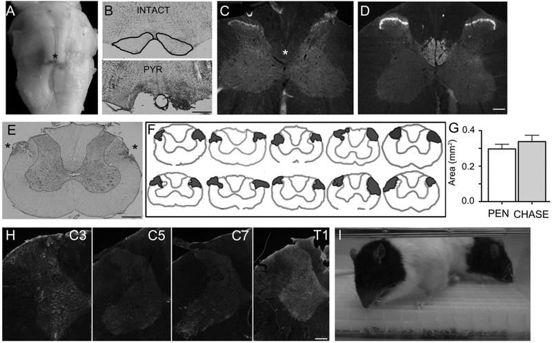
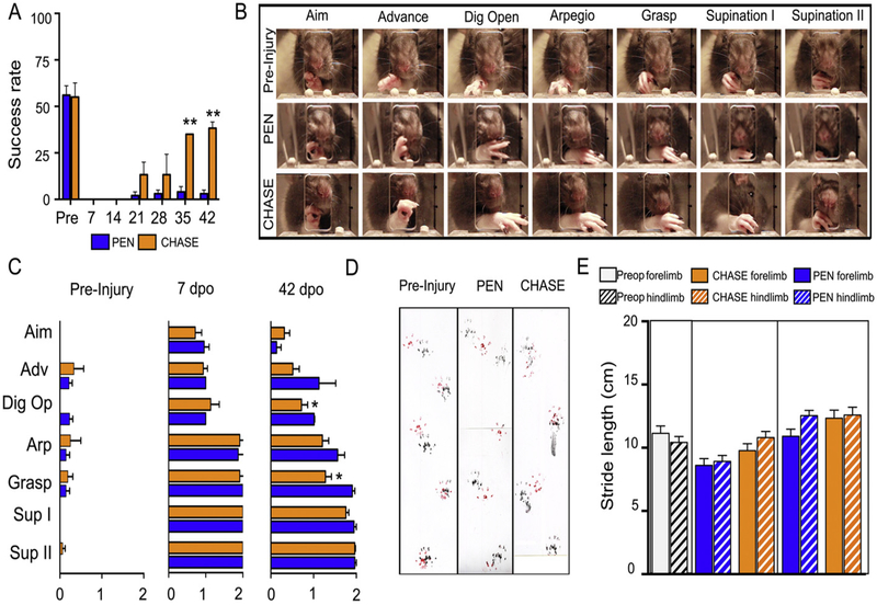
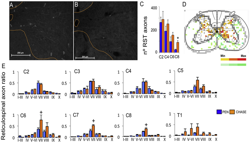
The corticospinal and rubrospinal tracts are the predominant tracts for controlling skilled hand function. Injuries to these tracts impair grasping but not gross motor functions such as overground locomotion. The aim of the present study was to determine whether or not, after damage to both the corticospinal and rubrospinal tracts, other spared subcortical motor pathway can mediate the recovery of skilled hand function. Adult rats received a bilateral injury to the corticospinal tract at the level of the medullar pyramids and a bilateral ablation of the rubrospinal axons at C4. One group of rats received, acutely after injury, two injections of chondroitinase-ABC at C7, and starting at 7days post-injury were enrolled in daily reaching and grasping rehabilitation (CHASE group, n=5). A second group of rats received analogous injections of ubiquitous penicillinase, and did not undergo rehabilitation (PEN group, n=5). Compared to rats in the PEN group, CHASE rats gradually recovered the ability to reach and grasp over 42days after injury. Overground locomotion was mildly affected after injury and both groups followed similar recovery. Since the reticulospinal tract plays a predominant role in motor control, we further investigated whether or not plasticity of this pathway could contribute to the animal's recovery. Reticulospinal axons were anterogradely traced in both groups of rats. The density of reticulospinal processes in both the normal and ectopic areas of the grey ventral matter of the caudal segments of the cervical spinal cord was greater in the CHASE than PEN group. The results indicate that after damage to spinal tracts that normally mediate the control of reaching and grasping in rats other complementary spinal tracts can acquire the role of those damaged tracts and promote task-specific recovery.

Keywords: Chondroitinase-ABC; Corticospinal; Plasticity; Reaching and grasping; Reticulospinal; Spinal cord injury.


Figures

Fig. 1.
Fig. 1.
(A) Ventral view of a brainstem with a bilateral injury to the pyramids (asterisk). (B) Transverse sections of the medullar pyramids of an intact (outlined in black) and pyramidotomized (PYR) rat. (C) Transverse sections from the C4 spinal segment of a pyramidotomized rat immunostained with γPKC, showing the absence of CST labeling (asterisk) compared to an uninjured spinal cord (D). Note the staining of neurons in Rexed laminae 1–3 in both the intact and injured rat, excluding the possibility of false negative staining. (E) Cresyl violet stained transverse section from the C4 spinal segment with a bilateral injury of the dorso-lateral funiculi (asterisks). (F) Illustration showing the contour of C4 spinal cord sections with the localization and extension of the bilateral dorsolateral funiculi injuries in individual rats. The top illustrations belong to CHASE animals and the bottom to PEN animals. (G) Bar graphs showing the mean (±SEM) transverse extension area of the bilateral dorsolateral funiculi injury for 5 rats in each group. (H) Transverse section of cervical spinal segments immunostained against CSPGs sugars with CS-56 antibody. The gray matter is outlined in orange. The tissues were processed in parallel and the images were taken under the same optical conditions. (I) Photograph of rats from the CHASE group inside a cage with a grid on the bottom filled with sunflower seeds undergoing reaching and grasping rehabilitation. Scale bars in B and E = 500 μm; in C, D, and H = 200 μm.
Fig. 2.
Fig. 2.
(A) Mean (±SEM) success rate for reaching and grasping performance for rats in the PEN and CHASE groups over 42 days post-injury. (B) Sequence of video frames showing each movement component during a reaching and grasping attempt previous to the injury and at 42 days post-injury in a representative rat in the PEN and CHASE groups (see Methods for description of each component). (C) Detailed behavioral analyses of the reaching and grasping components pre-injury and at 7 and 42 days post-injury (dpo). (D) Imprints of the forepaws (red) and hindpaws (black) of a rat pre-injury and from a representative rat in the PEN and CHASE groups at 42 days post-injury when stepping along a corridor. (E) Histograms showing the mean (±SEM) stride length bilaterally pre-injury and 7 and 42 days post-injury for all rats in the PEN and CHASE groups. ** P < 0.001 and *P < 0.01 vs. PEN.
Fig. 3.
Fig. 3.
Anterogradely traced reticulospinal processes in the ventral gray matter of the C7 spinal segment from a representative rat in the CHASE (A) and PEN (B) groups. (C) Mean (±SEM) total number of reticulospinal processes in the gray matter ispilateral to the injection side. (D) The number of processes was determined in 100 μm2 square grids and plotted as heat maps for each spinal segment. (E) Mean (±SEM) number of reticulospinal processes in each Rexed lamina of the spinal gray matter normalized to the number of reticulospinal axons in the white mater on the ispilateral side of the same spinal segment. +, P < 0.1 vs. PEN.* p < 0.05 vs PEN.

Similar articles

Cited by

References

    1. Alilain WJ, Horn KP, Hu H, Dick TE, Silver J, 2011. Functional regeneration of respiratory pathways after spinal cord injury. Nature 475, 196–200. - PMC - PubMed
    1. Alluin O, Delivet-Mongrain H, Gauthier MK, Fehlings MG, Rossignol S, Karimi-Abdolrezaee S, 2014. Examination of the combined effects of chondroitinase ABC, growth factors and locomotor training following compressive spinal cord injury on neuroanatomical plasticity and kinematics. PLoS One 9, e111072. - PMC - PubMed
    1. Alstermark B, Isa T, Lundberg A, Pettersson LG, Tantisira B, 1989. The effect of low pyramidal lesions on forelimb movements in the cat. Neurosci. Res 7, 71–75. - PubMed
    1. Bachmann LC, Lindau NT, Felder P, Schwab ME, 2014. Sprouting of brainstem-spinal tracts in response to unilateral motor cortex stroke in mice. J. Neurosci 34, 3378–3389. - PMC - PubMed
    1. Baker SN, 2011. The primate reticulospinal tract, hand function and functional recovery. J. Physiol 589, 5603–5612. - PMC - PubMed

Publication types