. 2015 Mar 20;290(12):7492-505.
doi: 10.1074/jbc.M114.614925. Epub 2015 Feb 9.

The E3 ubiquitin ligase parkin is recruited to the 26 S proteasome via the proteasomal ubiquitin receptor Rpn13

Affiliations

The E3 ubiquitin ligase parkin is recruited to the 26 S proteasome via the proteasomal ubiquitin receptor Rpn13

Miguel A Aguileta et al. J Biol Chem. .

Abstract

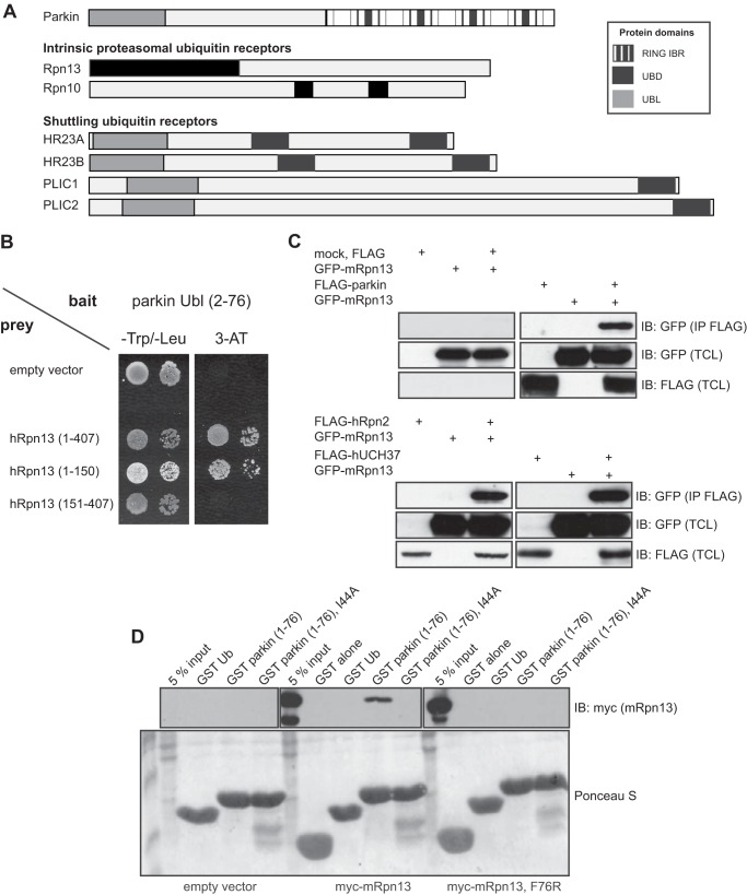
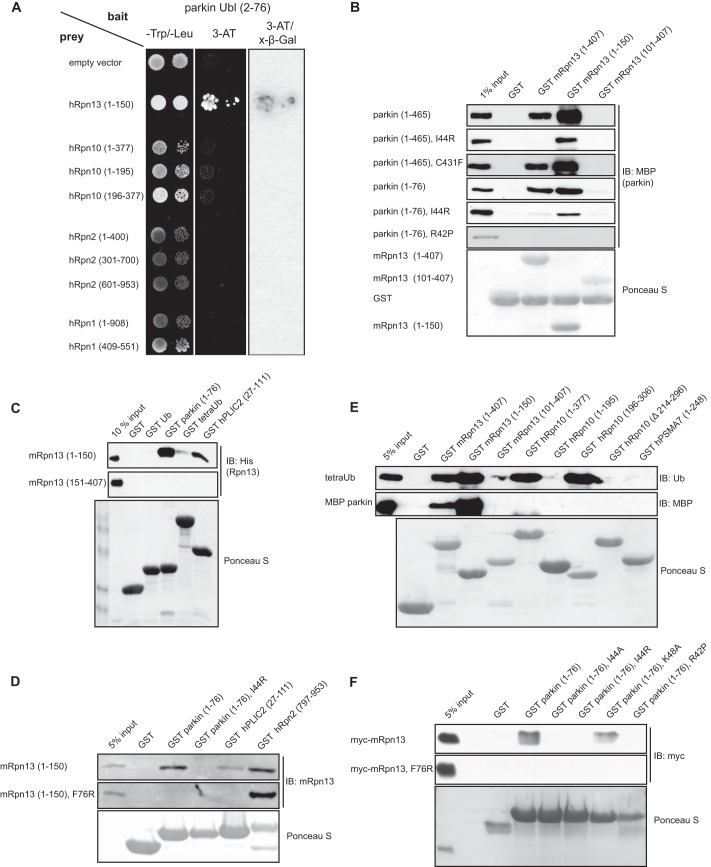
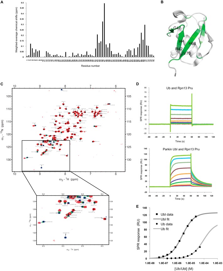
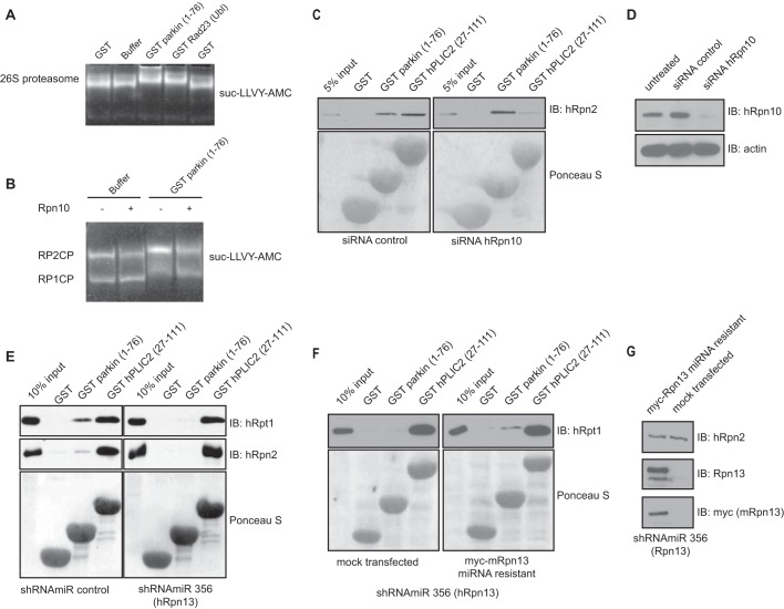
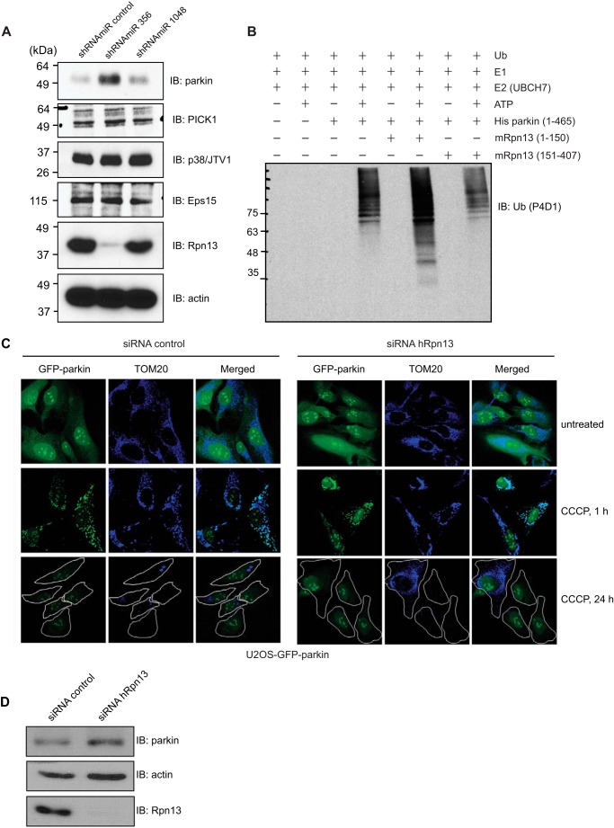
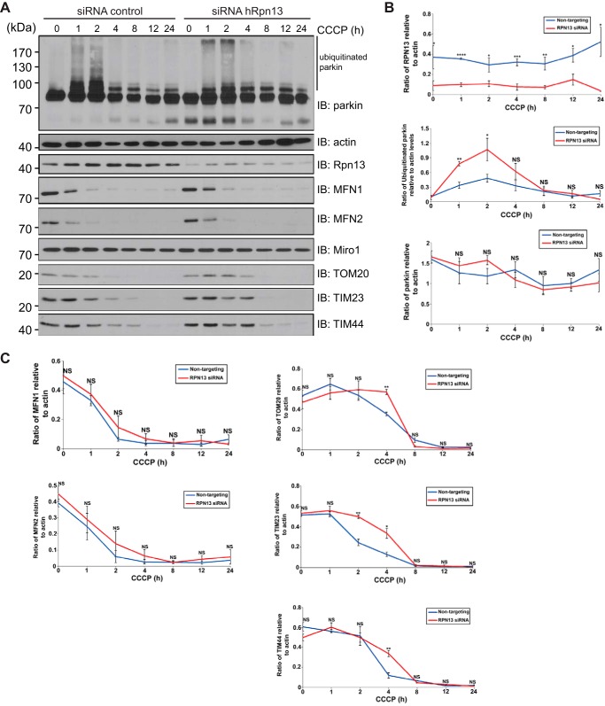
Mutations in the Park2 gene, encoding the RING-HECT hybrid E3 ubiquitin ligase parkin, are responsible for a common familial form of Parkinson disease. By mono- and polyubiquitinating target proteins, parkin regulates various cellular processes, including degradation of proteins within the 26 S proteasome, a large multimeric degradation machine. In our attempt to further elucidate the function of parkin, we have identified the proteasomal ubiquitin receptor Rpn13/ADRM1 as a parkin-interacting protein. We show that the N-terminal ubiquitin-like (Ubl) domain of parkin binds directly to the pleckstrin-like receptor for ubiquitin (Pru) domain within Rpn13. Using mutational analysis and NMR, we find that Pru binding involves the hydrophobic patch surrounding Ile-44 in the parkin Ubl, a region that is highly conserved between ubiquitin and Ubl domains. However, compared with ubiquitin, the parkin Ubl exhibits greater than 10-fold higher affinity for the Pru domain. Moreover, knockdown of Rpn13 in cells increases parkin levels and abrogates parkin recruitment to the 26 S proteasome, establishing Rpn13 as the major proteasomal receptor for parkin. In contrast, silencing Rpn13 did not impair parkin recruitment to mitochondria or parkin-mediated mitophagy upon carbonyl cyanide m-chlorophenyl hydrazone-induced mitochondrial depolarization. However, it did delay the clearance of mitochondrial proteins (TIM23, TIM44, and TOM20) and enhance parkin autoubiquitination. Taken together, these findings implicate Rpn13 in linking parkin to the 26 S proteasome and regulating the clearance of mitochondrial proteins during mitophagy.

Keywords: E3 Ubiquitin Ligase; Parkin; Parkinson Disease; Proteasome; Rpn13; Ubiquitin; Ubiquitylation (Ubiquitination).


Figures

FIGURE 1.
FIGURE 1.
The Ubl domain of parkin binds the Pru domain of Rpn13. A, schematic representation of the E3 ligase parkin and proteasome-related ubiquitin receptors. Specific domains and motifs are depicted. UBD, ubiquitin-binding domain; UBL, ubiquitin-like domain; IBR, in between ring fingers. B, mapping of the parkin Ubl and Rpn13 binding regions. Yeast strain Y190 was transformed with parkin Ubl-pYTH9 vector and various fragments of hRpn13 in pACT2 vector as preys. C, overexpressed mRpn13 and parkin bind, as shown by co-immunoprecipitation in HEK293T lysates. Plasmids for hRpn2 and hUCH37 were used as positive binding controls. D, residues involved in Rpn13-Ub binding also mediate the Rpn13-parkin interaction. Lysates from HEK293T cells transiently transfected with full-length Myc-mRpn13 (aa 1–407) or its corresponding F76R mutant were used for GST pulldown assays with GST alone, GST Ub (positive control), and either WT or I44A mutant of GST-tagged Ubl domain of parkin. Empty vector pcDNA 3.1 was used as a negative control. IB, immunoblotting; TCL, total cell lysate.
FIGURE 2.
FIGURE 2.
The Ubl domain of parkin interacts with the 19 S proteasomal subunit Rpn13. A, Ubl domain of parkin binds Pru domain of Rpn13. Yeast strain Y190 transformed with parkin Ubl-pYTH9 plasmid was additionally transformed with plasmids encoding various parts of mRpn13, hRpn10, hRpn2, and hRpn1 cDNA, and their binding was assayed by selective growth and X-β-Gal assay. B, Ubl domain of parkin directly binds Pru domain of Rpn13 via its Ile-44 hydrophobic patch. Using in vitro binding assay, various point and deletion mutants of parkin, expressed as MBP fusion proteins eluted from amylose resins, were tested for direct binding to different domains of Rpn13, which were expressed as GST fusion proteins and coupled to glutathione-Sepharose 4B beads. Ponceau S indicates levels of GST-coupled beads used in GST pulldown assay. Prior SDS-PAGE washed GST-beads were cleaved with thrombin for better visualization. C, Ubl domain of parkin, similar to ubiquitin and Ubl domain of hPLIC2 (aa 27–111), directly binds Pru domain of Rpn13. Using in vitro binding assay, ubiquitin and Ubl domains, expressed as GST fusion proteins, were tested for direct binding to different domains of Rpn13, which were expressed as His fusion proteins and eluted from nickel-nitrilotriacetic acid resins before GST pulldown assay. Ponceau S indicates levels of GST-coupled beads used in GST pulldown assay. D, loop mutation (F76R) in Rpn13 and hydrophobic patch mutation (I44R) in parkin abolish their direct binding. Purified WT (aa 1–150) and F76R Pru domain (aa 1–150), obtained by removal of GST moiety from GST fusion proteins by thrombin cleavage, were tested for binding to WT and I44R Ubl domain (aa 1–76) of parkin. Parkin constructs were expressed as GST-fused proteins coupled to glutathione-Sepharose 4B beads. As positive binding controls, GST-fused fragments of hRpn2 (aa 797–953) and hPLIC2 (aa 27–111) were used. E, purified MBP-fused parkin, eluted from amylose resins, was tested for direct binding to fragments of different proteasomal subunits, including mRpn13, hRpn10, and hPSMA7 as GST-tagged proteins. TetraUb cleaved with thrombin from GST beads served as a positive binding control. F, GST pulldown assay with lysates from HEK293T cells transiently transfected with full-length Myc-mRpn13 (upper panel) or F76R Myc-mRpn13 (lower panel) plasmids and GST alone (negative control) and various GST-fused mutants of parkin Ubl domain bound to beads. IB, immunoblotting.
FIGURE 3.
FIGURE 3.
NMR and SPR characterization of parkin Ubl binding to the Rpn13 Pru. A, weighted average NMR chemical shift perturbations in 15N-labeled parkin Ubl upon binding to Rpn13 Pru domain. B, NMR chemical shift perturbations mapped on the crystal structure of the murine parkin Ubl domain (Protein Data Bank code 2ZEQ (60)). Perturbations are colored in dark green (>0.15 ppm), medium green (0.1–0.15 ppm), or pale green (0.05–0.1 ppm). Important residues involved in the interaction are labeled in black. C, NMR 15N-1H heteronuclear single quantum coherence spectra of 15N-labeled parkin Ubl in the presence of Rpn13 Pru domain. Molar ratios were 0 (blue), 0.44 (green), 0.89 (purple), and 1.25 (red). Spectra are plotted at equivalent contour levels, and thresholds were adjusted to account for differences in protein concentrations and number of scans. The boxed area is enlarged below. Notice that there is significant broadening at 0.44 and 0.89 molar ratios for peaks that shift most (green and purple spectra). D, SPR sensorgrams of Ub or parkin Ubl analyte solution injected at different concentrations over immobilized ligand His6-tagged Rpn13 Pru. Notice the slower association and dissociation kinetics of the parkin Ubl compared with Ub. E, fitting of the SPR steady-state equilibrium responses as a function of analyte concentration to obtain equilibrium dissociation constants of 65 ± 25 and 3 ± 2 μm for Ub and Ubl for binding to His6-Rpn13 Pru.
FIGURE 4.
FIGURE 4.
Parkin interacts with the proteasome specifically via Rpn13. A, gel mobility shift assay of the interaction between the Ubl domain of parkin and the bovine proteasome. Gel mobility shift upon addition of GST (negative control), GST Rad23 Ubl (positive control), or GST parkin Ubl was monitored by nondenaturing PAGE and visualized with fluorogenic substrate suc-LLVY-AMC. Binding of the Ubl domain of parkin decreases the mobility of the proteasomes. B, gel mobility shift assay of the interaction between the Ubl domain of parkin and the proteasome purified from wild-type or Rpn10-deleted (Rpn10Δ) mutant of S. cerevisiae. The mobility of the proteasome following nondenaturing PAGE was visualized with fluorogenic substrate suc-LLVY-AMC. Binding of the Ubl domain of parkin decreased the mobility of the proteasomes irrespective of Rpn10 presence. RP1CP and RP2CP refer to singly or doubly 19 S capped 20 S proteasomes, respectively. Active proteasome was purified and analyzed by nondenaturating PAGE as described (61). C, a siRNA-mediated knockdown of hRpn10 does not affect parkin binding to proteasome. Lysates from HEK293T cells transfected with control nontargeting or anti hRpn10 siRNAs were incubated with GST, GST-parkin Ubl, and GST-hPLIC2 Ubl bound to glutathione-Sepharose 4B. Proteasome binding was assessed by immunoblotting against the 19 S proteasome subunit hRpn2. D, the efficiency of hRpn10 silencing in HEK293T cells (controls for C) was assessed by immunoblotting for hRpn10 and actin (as a loading control). E, silencing of Rpn13 abolishes parkin binding to the proteasome. Lysates from HEK293T cells infected with the indicated shRNAmiR were incubated with GST, GST-parkin Ubl, and GST-hPLIC2 Ubl bound to glutathione-Sepharose 4B. Proteasome binding was assessed by immunoblotting against the 19 S proteasome subunits hRpt1 and hRpn2. F, a miRNA-resistant form of Rpn13 rescues the interaction between parkin and the proteasome. shRNAmiR 356-infected HEK293T cells were transiently transfected with a miRNA-resistant Myc-Rpn13 construct or mock transfected. Binding of the parkin Ubl to the proteasome was detected by hRpt1 immunoblotting in lysates from cells transiently transfected with Myc-Rpn13, unlike in the mock-transfected cells. G, the efficiency of Rpn13 silencing and rescue with miRNA-resistant Myc-Rpn13 were assessed by immunoblotting for Rpn13, Myc, and hRpn2 (controls for A and B). IB, immunoblotting.
FIGURE 5.
FIGURE 5.
Interaction with Rpn13 increases the E3 ligase activity of parkin, whereas Rpn13 knockdown leads to increased level of parkin protein levels and has no effect on mitochondrial recruitment of parkin. A, parkin levels increase upon knockdown of Rpn13. Parkin protein levels only increase when shRNAmiR 356 and not the knockdown-ineffective shRNAmiR 1048 or control shRNAmiR are used. None of the analyzed parkin substrates (PICK1, p38/JTV1, and Eps15) were affected by the Rpn13 knockdown. Efficiency of Rpn13 silencing in HEK293T cells using lentivirus-delivered shRNAmiR against Rpn13 (shRNAmiR 356 and shRNAmiR 1048) and a control shRNAmiR was assessed by immunoblotting for Rpn13 and actin (as a loading control). B, in vitro ubiquitination reaction with purified His-parkin as E3 ligase. Reaction mixture (E1, E2 UbcH7, ATP, and ubiquitin) was complemented with equal amounts of various fragments of purified Rpn13. C, knockdown of Rpn13 has no effect on the mitochondrial recruitment of parkin or on the autophagic clearance of mitochondria. U2OS-GFP-parkin cells were transfected with nontargeting or Rpn13 siRNA (10 nm) for 60 h. Untreated cells or cells treated with CCCP for 1 or 24 h were fixed, and images were acquired after staining for the mitochondrial protein, TOM20. D, validation of Rpn13 siRNA knockdown. U2OS-GFP-parkin cells were transfected with nontargeting or Rpn13 siRNA oligonucleotides (10 nm) for 60 h. Cells were lysed and analyzed by immunoblotting for Rpn13, parkin, and actin. IB, immunoblotting.
FIGURE 6.
FIGURE 6.
Knockdown of Rpn13 delays the clearance of parkin substrates upon mitochondrial membrane depolarization. A, knockdown of Rpn13 delayed the clearance of TIM and TOM proteins. U2OS-GFP-parkin cells were transiently transfected with nontargeting or Rpn13 siRNA (10 nm) for 60 h. Cells were either left untreated or were treated with CCCP for the indicated intervals. Cells were lysed and analyzed by immunoblotting for parkin, Rpn13, actin, Mitofusins 1 and 2, Miro1, TIM23, TIM44, and TOM20 (quantified in Figs. 6B and 6C). B, quantification of Rpn13 and parkin protein levels upon CCCP treatment of Rpn13 knockdown U2OS-GFP-parkin cells. U2OS-GFP-parkin cells were transiently transfected with nontargeting or Rpn13 siRNA (10 nm) for 60 h and either left untreated or treated with CCCP for the indicated intervals (0–24 h). Protein levels of Rpn13 and ubiquitinated (parkin smear excluding unmodified parkin) and unmodified parkin upon CCCP treatment were compared between nontargeting and Rpn13 knockdown U2OS-GFP-parkin cells. The optical densities of the indicated proteins and total actin (used for normalization) were quantified using ImageJ (National Institutes of Health). The data represent the mean S.E. for three independent experiments. For statistical analysis, a two-way analysis of variance with Tukey post-test was performed. *, p < 0.05; **, p < 0.01; ***, p < 0.001; ****, p < 0.0001; NS, not significant. C, quantification of mitochondrial protein levels upon CCCP treatment of Rpn13 knockdown U2OS-GFP-parkin cells. U2OS-GFP-parkin cells were transiently transfected with nontargeting or Rpn13 siRNA (10 nm) for 60 h and either left untreated or treated with CCCP for the indicated intervals (0–24 h). Decrease in protein levels of MFN1, MFN2, TOM20, TIM23, and TIM44 upon CCCP treatment was compared between nontargeting and Rpn13 knockdown U2OS-GFP-parkin cells. Data analysis was performed as in Fig. 6B. IB, immunoblotting.

References

    1. Giasson B. I., Lee V. M. (2001) Parkin and the molecular pathways of Parkinson's disease. Neuron 31, 885–888 - PubMed
    1. Kitada T., Asakawa S., Hattori N., Matsumine H., Yamamura Y., Minoshima S., Yokochi M., Mizuno Y., Shimizu N. (1998) Mutations in the parkin gene cause autosomal recessive juvenile parkinsonism. Nature 392, 605–608 - PubMed
    1. Gasser T. (2009) Molecular pathogenesis of Parkinson disease: insights from genetic studies. Expert Rev. Mol. Med. 11, e22. - PubMed
    1. Shimura H., Hattori N., Kubo S., Mizuno Y., Asakawa S., Minoshima S., Shimizu N., Iwai K., Chiba T., Tanaka K., Suzuki T. (2000) Familial Parkinson disease gene product, parkin, is a ubiquitin-protein ligase. Nat. Genet. 25, 302–305 - PubMed
    1. Zhang Y., Gao J., Chung K. K., Huang H., Dawson V. L., Dawson T. M. (2000) Parkin functions as an E2-dependent ubiquitin-protein ligase and promotes the degradation of the synaptic vesicle-associated protein, CDCrel-1. Proc. Natl. Acad. Sci. U.S.A. 97, 13354–13359 - PMC - PubMed

Publication types

MeSH terms

Substances

LinkOut - more resources