Review
. 2015 May;148(6):1143-57.
doi: 10.1053/j.gastro.2015.02.002. Epub 2015 Feb 7.

Molecular, genetic, and cellular bases for treating eosinophilic esophagitis

Affiliations
Review

Molecular, genetic, and cellular bases for treating eosinophilic esophagitis

Marc E Rothenberg. Gastroenterology. 2015 May.

Abstract

Eosinophilic esophagitis (EoE) was historically distinguished from gastroesophageal reflux disease on the basis of histology and lack of responsiveness to acid suppressive therapy, but it is now appreciated that esophageal eosinophilia can respond to proton pump inhibitors. Genetic and environmental factors contribute to risk for EoE, particularly early-life events. Disease pathogenesis involves activation of epithelial inflammatory pathways (production of eotaxin-3 [encoded by CCL26]), impaired barrier function (mediated by loss of desmoglein-1), increased production and/or activity of transforming growth factor-β, and induction of allergic inflammation by eosinophils and mast cells. Susceptibility has been associated with variants at 5q22 (TSLP) and 2p23 (CAPN14), indicating roles for allergic sensitization and esophageal specific protease pathways. We propose that EoE is a unique disease characterized by food hypersensitivity; strong hereditability influenced by early-life exposures and esophageal-specific genetic risk variants; and allergic inflammation and that the disease is remitted by disrupting inflammatory and T-helper type 2 cytokine-mediated responses and through dietary elimination therapy.

Keywords: Allergy; Barrier; Cytokines; Diagnostics; Eosinophils; Epithelium; Esophagitis; Genetics; Therapy.


Figures

Figure 1
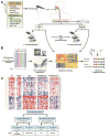
Figure 1. Diagnostic Evaluation of EoE, Including Next-generation Molecular Analysis
A, EoE should be considered for patients with characteristic symptoms of upper gastrointestinal diseases (note the age dependence) that are refractory to PPI treatment, and for patients with typical features of EoE (male, atopic, family history of esophageal dysfunction, and family history of EoE; note the high relative risk Ratio]. Patients with these features should undergo esophagogastroduodenoscopy; a diagnosis of EoE can be made for those with at least 15 eosinophils/hpf in esophageal biopsies. B, diagnosis and assessment of disease activity is facilitated by molecular analysis of esophageal tissue for the expression of 94 EoE-related transcripts (via the EoE Diagnostic Panel [EDP]). The EDP involves panel expression acquisition, signature analysis, and quantitative scoring for EoE-related processes (a Molecular Diagnostic Report). Recent attention has focused on analysis of PPI-responsive esophageal eosinophilia (PPI-REE), which has a transcript profile that overlaps with that of EoE but normalizes following PPI treatment. C, representative heat diagrams of the esophageal transcript profiles in EoE, PPI-REE before and after PPI therapy, as well as control normal (NL) and GERD patients. Figure is adapted from a recent publication.
Figure 2
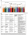
Figure 2. Genetic Associations in EoE
A, a Manhattan Plot with data from 736 subjects with EoE and 9246 controls (1,468,075 genetic variants) with minor allele frequencies greater than 1% in the subjects with EoE. The –log of the probability is shown as a function of the genomic position of the autosomes. Genome-wide significance (red dotted line, p ≤ 5×10⁻⁸) and suggestive significance (solid blue line, P≤ 10-7) are shown. The figure is adopted from a recent publication. B, a summary of the specific genes implicated in EoE susceptibility. The putative genes, the approach by which they were discovered, the type of identified genetic modifications, and plausible genetic mechanisms are listed. Mendelian inherited diseases associated with EoE are indicated.
Figure 3
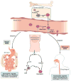
Figure 3. Eosinophil Development and Tissue Localization
Hematopoietic progenitor cells expand in response to FLT3 ligand (FLT3L) and stem cell factor (SCF); the eosinophil lineage is regulated by IL5 and the GATA1 transcription factor. IL5 subsequently promotes eosinophil migration from the bone marrow into the blood and maintains circulating levels of eosinophils. Eosinophil adhesion molecules (β7 and VLA4) interact with their respective endothelium receptors MAdCAM1 and VCAM1. Under baseline conditions, eosinophils migrate into all segments of the gastrointestinal tract (except the esophagus) in response to tissue eotaxin1. Intestinal eosinophils regulate sIgA production and intestinal microbiota composition. Innate lymphoid type 2 cells (ILC2) produce high levels of IL5 and IL13 in response to IL33 (via its receptor ST2). IL13 subsequently binds to its receptor IL13Rα1 on epithelial cells, which then produce eotaxin. In the esophagus, eosinophils accumulate in response to local production of eotaxin-3 and promote esophageal dysfunction and tissue remodeling.
Figure 4
Figure 4. Pathogenesis and Mechanism-based Therapy for EoE
A model of eosinophil development in the bone marrow, transit into the peripheral blood, trafficking to the esophagus, and the subsequent tissue changes, including impaired barrier function. In brief, allergens (aeroallergens and food allergens) induce Th2-associated inflammation, with a primary IL13-induced tissue response that involves modification of an extensive set of esophageal transcripts, including eotaxin3, which recruits eosinophils. Impaired barrier function is mediated by a pathogenic cycle induced by IL13 that results in decreased expression of desmoglein-1 (DSG1) (also associated with SAM) and subsequent induction of periostin. These changes promote tissue remodeling and eosinophil adhesion, as well as production of TSLP, which propagates the allergic inflammatory cascade. Local IgE and IgG4 are produced and mast cells are sensitized. Certain genetic elements increase susceptibility to EoE, including 2p23 (CAPN14), 5q22 (TSLP), and genes encoding the TSLP receptor and filaggrin. Their expression is modulated by miR-21 and miR-223. Therapies for EoE include PPI agents, topical glucocorticoids, elimination diets, and anti-cytokine humanized antibodies (e.g. anti-IL5, anti-IL13, anti-IL4Rα).
Figure 5
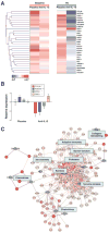
Figure 5. Molecular Evidence for the Efficacy of Anti-IL13 and Identification of IL13-associated Molecular Nodes
Transcriptome profile of esophageal biopsies at baseline and following 3 months of treatment with humanized anti-IL13 or placebo. A shows the heat map of the top differentially modified genes. B shows the the quantitative levels of representative genes. Note the increased expression of genes involved in eosinophil chemoattraction (CCL26), tissue remodeling (POSTN), mast cell activity (CPA3), and barrier dysfunction (DSG1). C, the comprehensive pathways, focused on protein-protein interactions, shows the in vivo IL13 networks. Several communication centers (shown as blue nodes) contribute to most of the organization and regulating functions. The different levels of red indicate the statistical strength of the interactions. Figures are adapted from a recent publication.

References

    1. Liacouras CA, Furuta GT, Hirano I, et al. Eosinophilic esophagitis: updated consensus recommendations for children and adults. J Allergy Clin Immunol. 2011;128:3–20 e6. quiz 21-2. - PubMed
    1. Dellon ES, Gonsalves N, Hirano I, et al. ACG Clinical Guideline: Evidenced Based Approach to the Diagnosis and Management of Esophageal Eosinophilia and Eosinophilic Esophagitis (EoE) Am J Gastroenterol. 2013;108:679–92. - PubMed
    1. Dellon EP, Liacouras C. Eosinophilic Esophagitis. Gastroenterol. 2015 In Press.
    1. Attwood SE, Smyrk TC, Demeester TR, et al. Esophageal eosinophilia with dysphagia. A distinct clinicopathologic syndrome. Dig Dis Sci. 1993;38:109–16. - PubMed
    1. Kelly KJ, Lazenby AJ, Rowe PC, et al. Eosinophilic esophagitis attributed to gastroesophageal reflux: improvement with an amino acid-based formula. Gastroenterology. 1995;109:1503–12. - PubMed

Publication types

MeSH terms