. 2015 May;21(5):435-51.
doi: 10.1093/molehr/gav006. Epub 2015 Feb 9.

Epidermal growth factor-like domain 7 promotes migration and invasion of human trophoblast cells through activation of MAPK, PI3K and NOTCH signaling pathways

Affiliations

Epidermal growth factor-like domain 7 promotes migration and invasion of human trophoblast cells through activation of MAPK, PI3K and NOTCH signaling pathways

M Massimiani et al. Mol Hum Reprod. 2015 May.

Abstract

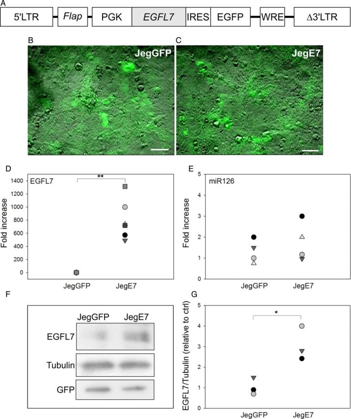
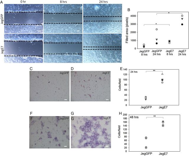
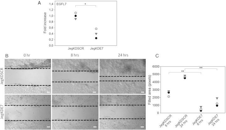
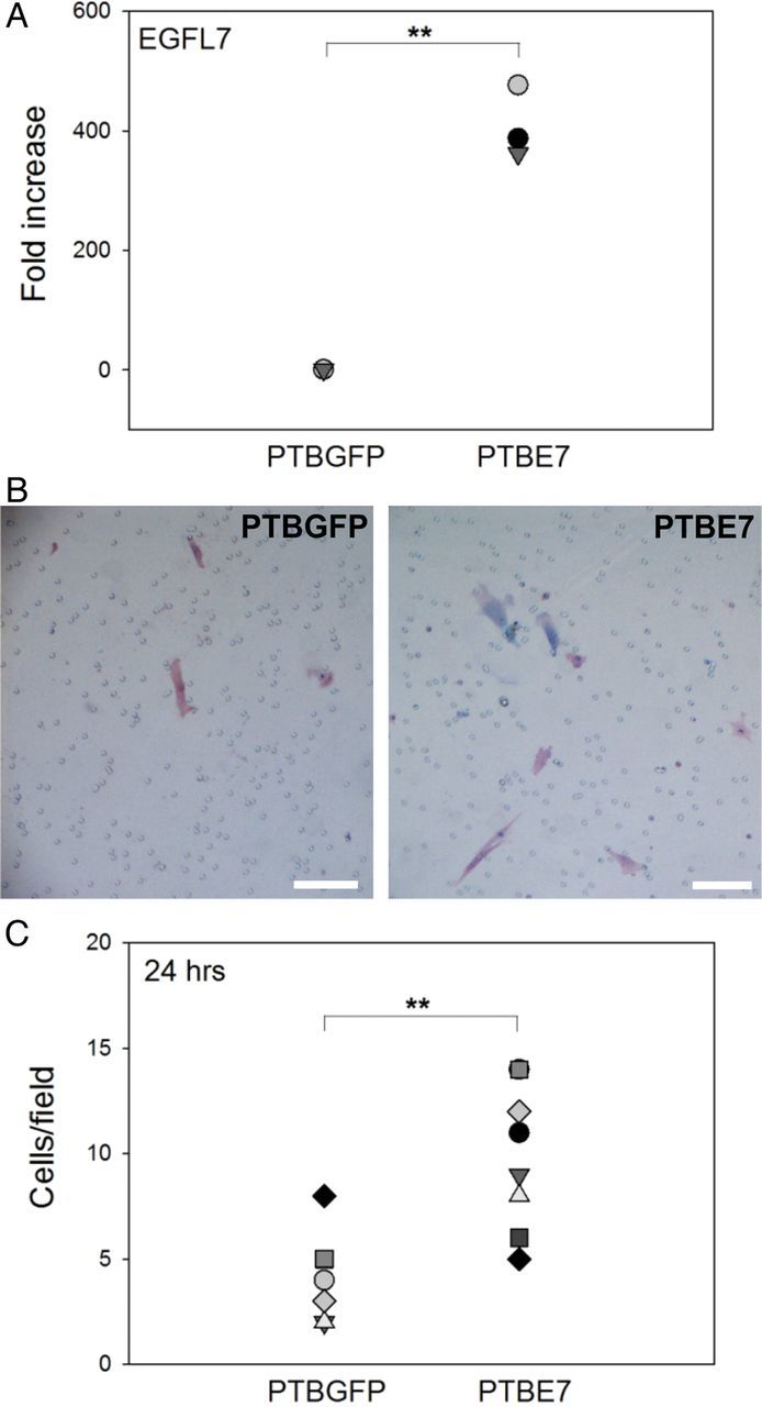
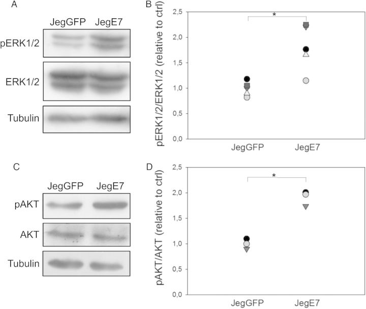
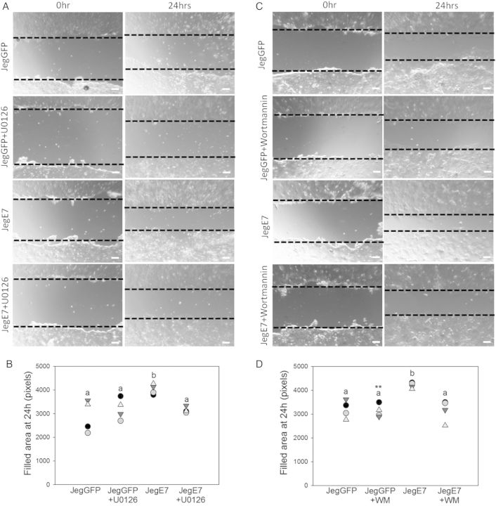
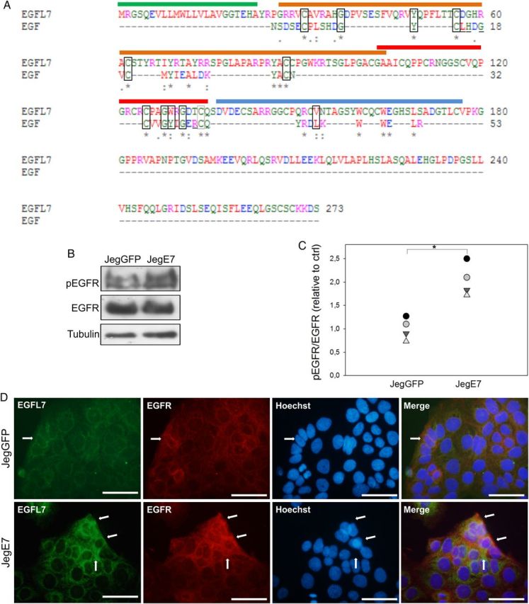
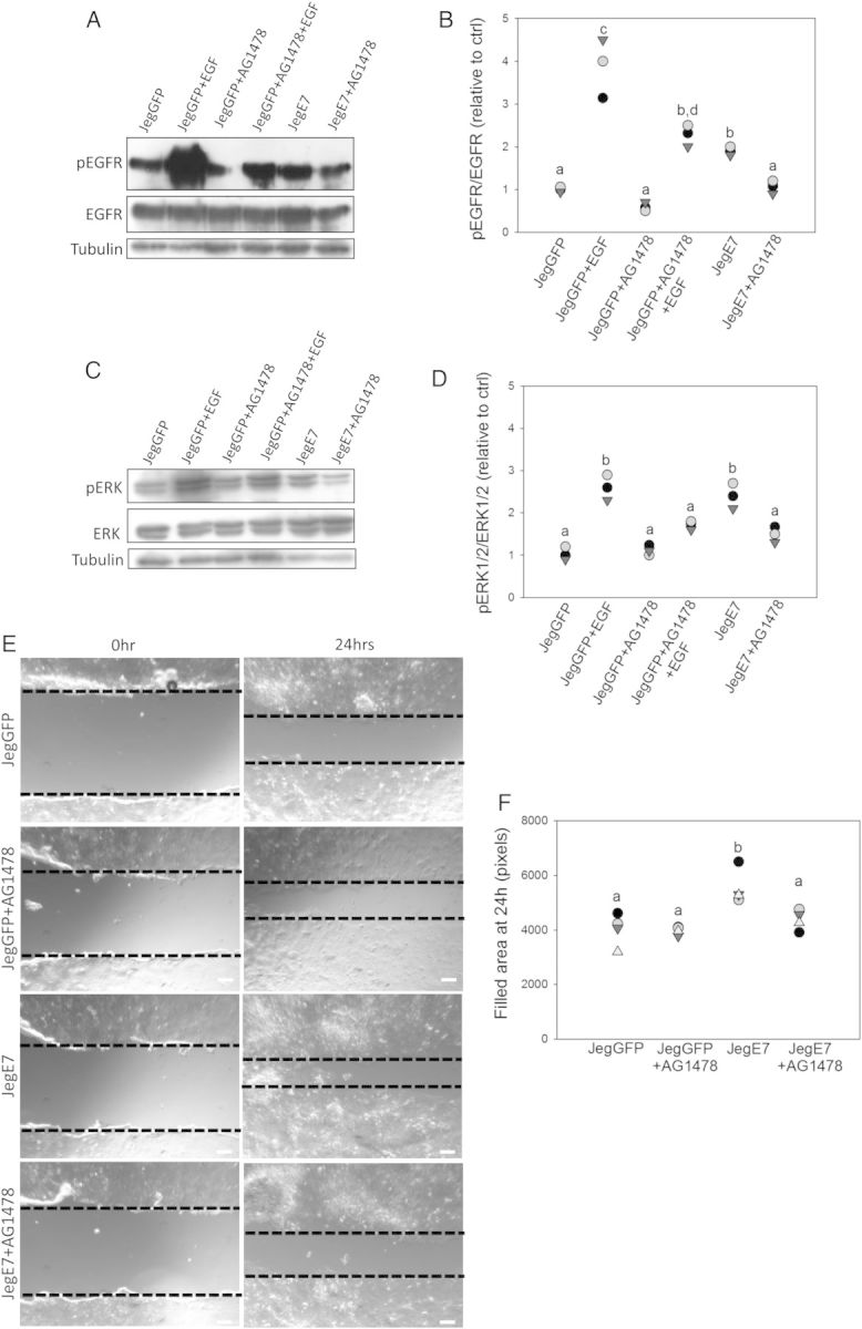
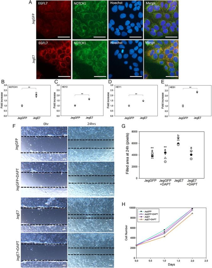
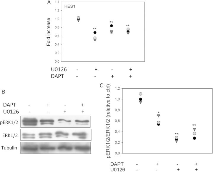
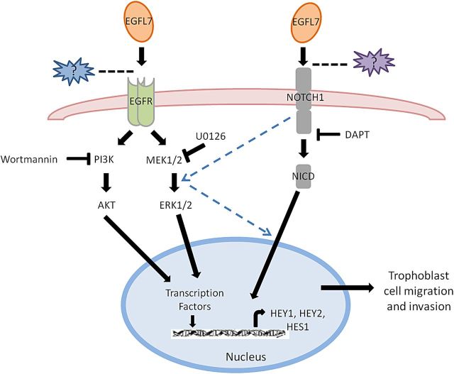
Epidermal growth factor-like domain 7 (Egfl7) is a gene that encodes a partially secreted protein and whose expression is largely restricted to the endothelia. We recently reported that EGFL7 is also expressed by trophoblast cells in mouse and human placentas. Here, we investigated the molecular pathways that are regulated by EGFL7 in trophoblast cells. Stable EGFL7 overexpression in a Jeg3 human choriocarcinoma cell line resulted in significantly increased cell migration and invasiveness, while cell proliferation was unaffected. Analysis of mitogen-activated protein kinase (MAPK) and phosphatidylinositol 3-kinase (PI3K) pathways showed that EGFL7 promotes Jeg3 cell motility by activating both pathways. We show that EGFL7 activates the epidermal growth factor receptor (EGFR) in Jeg3 cells, resulting in downstream activation of extracellular regulated kinases (ERKs). In addition, we provide evidence that EGFL7-triggered migration of Jeg3 cells involves activation of NOTCH signaling. EGFL7 and NOTCH1 are co-expressed in Jeg3 cells, and blocking of NOTCH activation abrogates enhanced migration of Jeg3 cells overexpressing EGFL7. We also demonstrate that signaling through EGFR and NOTCH converged to mediate EGFL7 effects. Reduction of endogenous EGFL7 expression in Jeg3 cells significantly decreased cell migration. We further confirmed that EGFL7 stimulates cell migration by using primary human first trimester trophoblast (PTB) cells overexpressing EGFL7. In conclusion, our data suggest that in trophoblast cells, EGFL7 regulates cell migration and invasion by activating multiple signaling pathways. Our results provide a possible explanation for the correlation between reduced expression of EGFL7 and inadequate trophoblast invasion observed in placentopathies.

Keywords: EGFL7; MAPK; NOTCH signaling; PI3K; trophoblast migration.


Figures

Figure 1
Figure 1
Generation of Jeg3 cells overexpressing epidermal growth factor-like domain 7 (EGFL7). (A) Schematic representation of lentiviral vector containing the human EGFL7 cDNA, an internal ribosome entry site (IRES) and a green fluorescent protein (GFP) reporter gene under the control of a phosphoglycerate kinase 1 (PGK) promoter. (B and C) GFP-positive Jeg3 cells transduced with the empty vector control (JegGFP) (B) and containing an EGFL7 cDNA (JegE7) (C). (D and E) Quantitative RT–PCR data for EGFL7 (D) and miR126 (E) in JegGFP and JegE7 cells. *P < 0.05; **P < 0.001. (FG) Western blot analysis for EGFL7 (F) and its densitometric analysis (G). Scale bars = 50 µm.
Figure 2
Figure 2
Epidermal growth factor-like domain 7 (EGFL7) stimulates migration and invasion of Jeg3 cells. (A) Wounding assay at 0, 8 and 24 h. Dotted lines indicate the edges of the monolayer. (B) Quantification of Jeg3 migration in wounding assay; the dot plots represent the area filled after 8 and 24 h. (CE) JegGFP (control) (C) and JegE7 (overexpressing EGFL7) (D) cells migrated under the filter of a transwell chamber after 24 h of culture. Quantification of cell migration is shown in the dot plot graph (E). (FH) JegGFP (F) and JegE7 (G) that invaded into the Matrigel and migrated under the filter of the transwell chamber after 48 h of culture. Quantification of cell invasion is shown in the dot plot graph (H). All data are represented as cells counted in each of at least six different fields for each experiment. *P < 0.05, **P < 0.001. Scale bars (A) = 120 µm; (C;D;F;G) = 60 µm.
Figure 3
Figure 3
Epidermal growth factor-like domain 7 (EGFL7) knockdown reduces migration of Jeg3 cells. (A) Quantitative RT–PCR data for EGFL7 in scrambled control (JegKDSCR) and EGFL7 knockdown Jeg3 cells (JegKDE7). (B) Wounding assay at 0, 8 and 24 h. Dotted lines indicate the edges of the monolayer. (C) Quantification of Jeg3 migration in wounding assay, represented as dot plot showing the area filled after at 8 and 24 h. *P < 0.05, **P < 0.001. Scale bars = 120 µm.
Figure 4
Figure 4
Generation of long-term primary human first trimester trophoblast (PTB) cells overexpressing epidermal growth factor-like domain 7 (PTBE7). EGFL7 stimulates migration of PTB cells. (A) Quantitative RT–PCR data for EGFL7. (C and B) PTBGFP (control) and PTBE7 cells migrated under the filter of transwell chamber after 24 h of culture. Quantification of cell migration is shown in the dot plot graph (F). **P < 0.001. Scale bar (A-B) = 50 µm; (D-E) = 100 µm.
Figure 5
Figure 5
Epidermal growth factor-like domain 7 (EGFL7) activates mitogen-activated protein kinase (MAPK) and phosphatidylinositol 3-kinase (PI3K) pathways. Western blot analysis of cell extracts from JegGFP (control) and JegE7 (overexpressing EGFL7) cells for phosphorylated extracellular regulated kinase 1 and 2 (pERK1/2) (A) and phosphorylated protein kinase B (pAKT) (C) in JegGFP and JegE7, with Tubulin as a loading control. The respective densitometric analysis (B and D). Data are presented as percentage of control. *P < 0.05.
Figure 6
Figure 6
Epidermal growth factor-like domain 7 (EGFL7) promotes Jeg3 cell migration through mitogen-activated protein kinase (MAPK) and phosphatidylinositol 3-kinase (PI3K) signaling pathways. Dotted lines highlight the edges of the monolayer. (A) Wounding assay at 0 and 24 h with or without addition of the mitogen-activated protein/extracellular signal-regulated kinase kinase (MEK 1/2)-inhibitor U0126 (5 µM). (B) Quantification of Jeg3 migration in wounding assay, represented as dot plot, showing the total area filled at 24 h. (C) Wounding assay at 0 and 24 h with or without addition of the PI3K-inhibitor Wortmannin (100 nM). (D) Quantification of Jeg3 migration in wounding assay, represented as dot plot showing the total area filled at 24 h. Different letters are to be considered within each culture time-point and indicate statistically significant differences. P < 0.001 is indicated by **. Scale bars = 120 µm. WM = Wortmannin.
Figure 7
Figure 7
Epidermal growth factor-like domain 7 (EGFL7) interacts with and activates epidermal growth factor receptor (EGFR) in Jeg3 cells. (A) Alignment of the amino acid (aa) sequences of EGFL7 and epidermal growth factor (EGF). The signal peptide of EGFL7 is marked with a green line (aa 1–23), the EMI domain with an orange line (aa 27–104), the EGF-like domain with a red line (aa 103–135), the Ca2+ EGF-like domain with a blue line (aa 137–177). Amino acids involved in EGFR binding that are conserved between EGF and EGFL7 are depicted by rectangles. Amino acids that are involved in EGFR binding but are not conserved between EGF and EGFL7 are underlined (Van Zoelen et al., 2000). Asterisks = positions which have a single, fully conserved residue; colons = conservation between groups of strongly similar properties; periods = conservation between groups of weakly similar properties. Legend for the colors: Red = AVFPMILW, small, hydrophobic; Blue = DE, acidic; Magenta = RK, basic; Green = STYHCNGQ, hydroxyl, sulfhydryl, amine; Grey = others, unusual aa. (B) Western blot analysis of pEGFR in JegGFP (control) and JegE7 (overexpressing EGFL7), with densitometric analysis (C). (D) Double immunofluorescent staining of JegGFP and JegE7 cells for EGFL7 (green), EGFR (red), and Hoechst (blue). Arrows indicate co-localization of EGFL7 and EGFR. *P < 0.05. Scale bars = 50 µm.
Figure 8
Figure 8
Epidermal growth factor-like domain 7 (EGFL7) activates the mitogen-activated protein kinase (MAPK) signaling pathway and promotes cell migration through epidermal growth factor receptor (EGFR). Jeg3 cells were treated with the EGFR-inhibitor AG1478 (1 µM) in serum-free culture medium for 30 min and/or stimulated with 10 ng/ml epidermal growth factor (EGF) for 30 min consecutively. Phosphorylation levels of EGFR (A) and extracellular regulated kinase 1 and 2 (ERK1/2) (C) were analyzed by western blot analysis. Respective densitometric analysis for (A and C) is shown in (B and D). (E) Wounding assay at 0 and 24 h in the presence of AG1478 (1 µM). Dotted lines highlight the edges of the monolayer. (F) Quantification of Jeg3 migration in the wounding assay, represented as dot plot showing the area filled at 24 h. Different letters are to be considered within each culture time-point and indicate statistically significant differences (P < 0.05). Scale bars = 120 µm.
Figure 9
Figure 9
Epidermal growth factor-like domain 7 (EGFL7) promotes Jeg3 cell migration also through NOTCH signaling pathway. (A) Double immunofluorescent staining of JegGFP (control) and JegE7 (overexpressing EGFL7) cells for EGFL7 (red), NOTCH1 (green), and Hoechst (blue). (BE) Quantitative RT–PCR data for transcript level of NOTCH1 (B), hairy and enhancer of split-related protein 2 (HEY-2) (C), hairy and enhancer of split-related protein 1 (HEY-1) (D), and hairy and enhancer of split-1 (HES-1) (E) in JegGFP and JegE7 cells. (F) Wounding assay at 0 and 24 h in presence of the γ-secretase inhibitor dual anti-platelet therapy (DAPT) (10 µM). Dotted lines highlight the edges of the monolayer. (G) Quantification of Jeg3 migration in wounding assay, represented as dot plot showing the area filled after 24 h. (H) JegGFP and JegE7 proliferation in the presence of DAPT (10 µM) was measured over 2 consecutive days by using the colorimetric 2-(4-iodophenyl)-3-(4-nitrophenyl)-5-(2,4-disulfophenyl)-2H-tetrazolium, monosodium salt (WST-1) assay; n = 4. Different letters are to be considered within each culture time-point and indicate statistically significant differences (P < 0.05); **P < 0.001. Scale bars (A) = 50 µm; (F) = 120 µm.
Figure 10
Figure 10
Inhibition of extracellular regulated kinase 1 and 2 (ERK1/2) activation reduces NOTCH signaling and blockage of NOTCH activity inhibits mitogen-activated protein kinase (MAPK) signaling pathway. (A) JegE7 (overexpressing EGFL7) cells were treated with the mitogen-activated protein/extracellular signal-regulated kinase kinase (MEK 1/2)-inhibitor U0126 (10 µM) or/and the γ-secretase inhibitor dual anti-platelet therapy DAPT (25 µM) in serum-free culture medium for 90 min. Transcript level of hairy and enhancer of split-1 (HES1) was analyzed by quantitative RT–PCR. (B and C) JegE7 cells were treated with DAPT (25 µM) or/and U0126 (10 µM) in serum-free culture medium for 60 min. Phosphorylation levels of ERK1/2 (B) were analyzed by western blot analysis. Respective densitometric analysis is shown in (C). *P < 0.05, **P < 0.001.
Figure 11
Figure 11
Schematic representation of cell signaling pathways involved in epidermal growth factor-like domain 7 (EGFL7)-induced trophoblast migration and invasion. Based on our findings, we propose that EGFL7 regulates trophoblast migration/invasion through the activation of the epidermal growth factor receptor (EGFR) and NOTCH pathways. The two pathways converge to mediate EGFL7 effects, as represented by the dashed lines. Question marks indicate possible other molecules mediating the interaction between EGFL7 and either EGFR or NOTCH1.

Similar articles

Cited by

References

    1. Bambino K, Lacko LA, Hajjar KA, Stuhlmann H. Epidermal growth factor-like domain 7 is a marker of the endothelial lineage and active angiogenesis. Genesis 2014;52:657–670. - PMC - PubMed
    1. Campagnolo L, Leahy A, Chitnis S, Koschnick S, Fitch MJ, Fallon JT, Loskutoff D, Taubman MB, Stuhlmann H. EGFL7 is a chemoattractant for endothelial cells and is up-regulated in angiogenesis and arterial injury. Am J Pathol 2005;167:275–284. - PMC - PubMed
    1. Campagnolo L, Moscatelli I, Pellegrini M, Siracusa G, Stuhlmann H. Expression of EGFL7 in primordial germ cells and in adult ovaries and testes. Gene Expr Patterns 2008;8:389–396. - PMC - PubMed
    1. Chakraborty C, Barbin YP, Chakrabarti S, Chidiac P, Dixon SJ, Lala PK. Endothelin-1 promotes migration and induces elevation of [ca2+]i and phosphorylation of map kinase of a human extravillous trophoblast cell line. Mol Cell Endocrinol 2003;201:63–73. - PubMed
    1. Cobellis L, Mastrogiacomo A, Federico E, Schettino MT, De Falco M, Manente L, Coppola G, Torella M, Colacurci N, De Luca A. Distribution of Notch protein members in normal and preeclampsia-complicated placentas. Cell Tissue Res 2007;330:527–534. - PubMed

Publication types

MeSH terms