. 2015 Mar;9(3):697-706.
doi: 10.3892/etm.2015.2185. Epub 2015 Jan 16.

Naringenin suppresses K562 human leukemia cell proliferation and ameliorates Adriamycin-induced oxidative damage in polymorphonuclear leukocytes

Affiliations

Naringenin suppresses K562 human leukemia cell proliferation and ameliorates Adriamycin-induced oxidative damage in polymorphonuclear leukocytes

Rui-Fang Li et al. Exp Ther Med. 2015 Mar.

Abstract

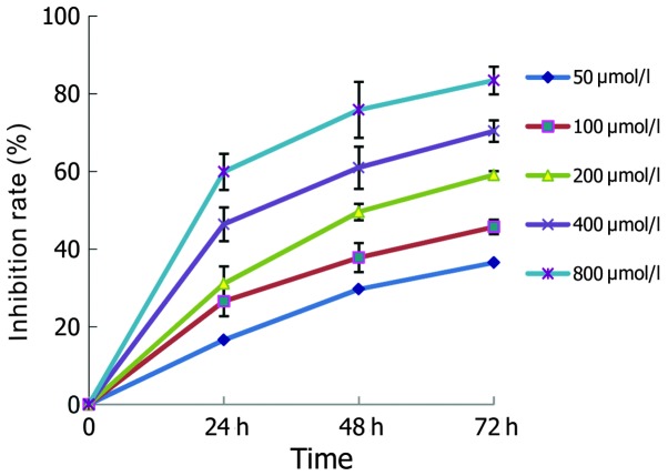
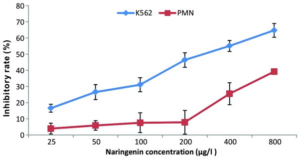
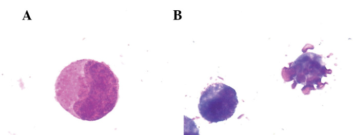
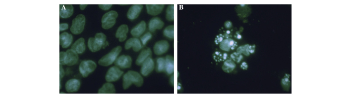
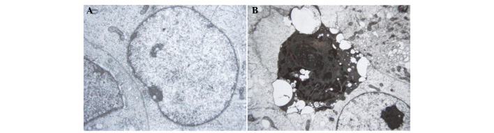
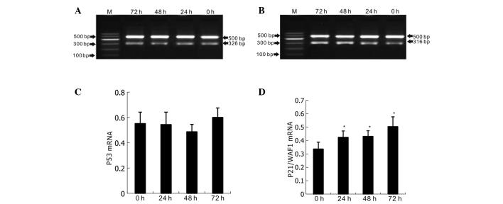
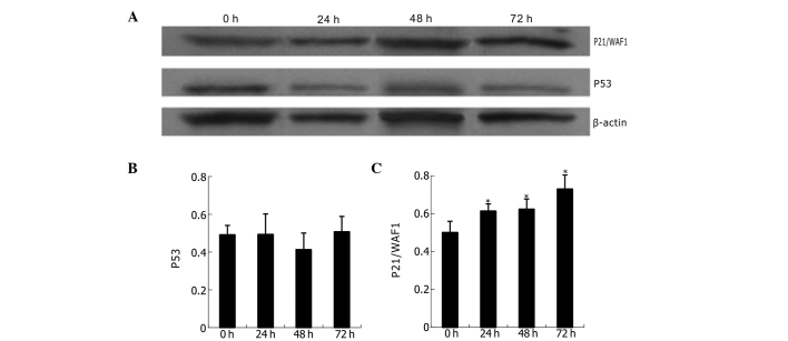
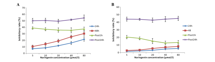
Treatments for leukemia remain unsatisfactory. Conventional chemotherapy agents that aim to kill tumor cells may also damage normal cells and thus result in severe side-effects. Naringenin, a natural polyphenolic compound with antioxidant effects, has been revealed to have significant antitumor effects with low toxicity in preliminary studies. Thus, it is considered as one of the most promising flavonoids in the treatment of leukemia. In the present study, the effects of naringenin on the K562 human leukemia cell line and the underlying mechanisms were explored in vitro. In addition, human peripheral blood polymorphonuclear leukocytes (PMNs) were used as a normal control in order to evaluate the effects of naringenin on normal granulocytes and in the mediation of Adriamycin (ADM)-induced oxidative damage. The results revealed that K562 proliferation was significantly inhibited by naringenin in a time- and concentration-dependent manner; however, minimal cytotoxic effects were observed in PMNs when naringenin was used at concentrations <400 μmol/l. Morphological changes indicative of apoptosis were observed in naringenin-treated K562 cells. Flow cytometric analysis indicated that the K562 cells were arrested in the G0/G1 phase of the cell cycle with a significantly upregulated rate of apoptosis. Furthermore, in the naringenin-treated K562 cells, the labeling index of proliferating cell nuclear antigen was observed to be increased by immunochemical staining, the mRNA and protein expression levels of p21/WAF1 were strongly upregulated in reverse transcription-polymerase chain reaction and western blot analyses, whereas p53 gene expression was not significantly changed. In PMNs to which naringenin (50~80 μmol/l) was added 1 h subsequent to ADM, the cell damage induced by ADM was significantly reduced, coincident with reductions in the levels of reactive oxygen species (ROS) and malondialdehyde (MDA) and increases in the activity of superoxide dismutase and glutathione peroxidase. However, the cytotoxic effect of ADM in K562 cells was not significantly altered by naringenin, and the oxidative stress indices in K562 cells remained stable. In conclusion, the present study revealed the promising value of naringenin in leukemia treatment. Naringenin demonstrated a significant inhibitory effect on the growth of K562 cells but not on normal PMNs. Furthermore, naringenin protected PMNs from ADM-induced oxidative damage at low concentrations. Cell cycle arrest and apoptosis-inducing effects, achieved through p53-independent p21/WAF1 upregulation, are likely to be the mechanism of the antileukemic effects of naringenin, and the protective effect against ADM chemotherapy-induced damage in PMNs may be due to the antioxidant capability of this agent at low concentrations.

Keywords: K562; apoptosis; cell cycle; cell line; gene; naringenin; p21; p53.


Figures

Figure 1
Figure 1
Growth inhibitory effect of naringenin on K562 cells.
Figure 2
Figure 2
Effects of naringenin on the growth of K562 cells and PMNs (48 h). PMN, polymorphonuclear leukocyte.
Figure 3
Figure 3
Morphologic observation of K562 cells with Wright-Giemsa staining. Cell morphology of (A) K562 cells in the control group and (B) K562 cells in the naringenin-treated group, which indicates the formation of apoptotic bodies. Magnification, ×1,000.
Figure 4
Figure 4
Morphological changes in K562 cells following naringenin treatment as revealed by confocal microscopy and Hoechst 33258 fluorescence staining. (A) K562 cells in the control group. (B) K562 cells treated with 400 μmol/l naringenin treatment for 24 h, which indicates morphological changes typical of apoptosis and the formation of apoptotic bodies. Magnification, ×400.
Figure 5
Figure 5
Ultrastructural changes of K652 cells with TEM observation. (A) Ultrastructure of K562 cells in the control group. TEM shows a smooth cell body, evenly dispersed chromatin and complete membrane and nuclear membrane. (B) K562 cells treated with 400 μmol/l of naringenin for 24 h displaying morphological changes typical of apoptosis, including cell nuclear condensation, nuclear membrane rupture, the formation of clumps along the nuclear membrane or crescent formation, and cytoplasm blistering. Magnification, ×15,000. TEM, transmission electron microscopy.
Figure 6
Figure 6
Semi-quantitative RT-PCR testing of p53 and p21/WAF1 expression in K562 cells with 400 μmol/l naringenin treatment. (A) and (B) electrophoresis of RT-PCR for p53 and p21 mRNA, respectively. M represents marker; bands from bottom to top indicate 100, 200, 300, 400, 500 and 600 bp in the ladder, respectively. β-actin (500 bp band) was used as an internal standard. The target gene fragment of p53 is 326 bp and of p21/WAF1 is 316 bp. (C) and (D) Bar charts of the relative content analysis of p53 and p21/WAF1 mRNA, respectively. Naringenin was used at a dose of 400 μmol/l. *P<0.05 vs. 0 h.
Figure 7
Figure 7
Western blots of p53 and p21/WAF1 protein in K562 cells treated with naringenin. (A) Western blot analysis of p53 and p21/WAF1 protein expression in K562 cells; β-actin was used as internal standard. (B) and (C) Bar charts of the relative content analysis of p53 and p21/WAF1, respectively, which were calculated by comparing the target gene to the internal reference β-actin. Naringenin was used at a dose of 400 μmol/l. *P<0.05 vs. 0 h.
Figure 8
Figure 8
Efficacy of naringenin in modulating the chemotherapeutic injury exerted by ADM on PMNs and K562 cells. Effects on (A) K562 cells and (B) PMNs. ADM, Adriamycin; PMN, polymorphonuclear leukocyte.

References

    1. Yang C, Zhang X. Incidence survey of leukemia in China. Chin Med Sci J. 1991;6:65–70. - PubMed
    1. Friesen C, Herr I, Krammer PH, Debatin KM. Involvement of the CD95 (APO-1/FAS) receptor/ligand system in drug-induced apoptosis in leukemia cells. Nat Med. 1996;2:574–577. doi: 10.1038/nm0596-574. - DOI - PubMed
    1. Durgo K, Koncar M, Komes D, et al. Cytotoxicity of blended versus single medicinal mushroom extracts on human cancer cell lines: contribution of polyphenol and polysaccharide content. Int J Med Mushrooms. 2013;15:435–448. doi: 10.1615/IntJMedMushr.v15.i5.20. - DOI - PubMed
    1. Ravishankar D, Rajora AK, Greco F, Osborn HM. Flavonoids as prospective compounds for anti-cancer therapy. Int J Biochem Cell Biol. 2013;45:2821–2831. doi: 10.1016/j.biocel.2013.10.004. - DOI - PubMed
    1. Ameer B, Weintraub RA, Johnson JV, Yost RA, Rouseff RL. Flavanone absorption after naringin, hesperidin, and citrus administration. Clin Pharmacol Ther. 1996;60:34–40. doi: 10.1016/S0009-9236(96)90164-2. - DOI - PubMed

LinkOut - more resources