

Central and peripheral corneal thickness measurement in normal and keratoconic eyes using three corneal pachymeters
- PMID: 25667728
- PMCID: PMC4307658
- DOI: 10.4103/2008-322X.143356
Central and peripheral corneal thickness measurement in normal and keratoconic eyes using three corneal pachymeters
Abstract
Purpose: To assess the agreement among ultrasonic pachymetry, the Galilei dual Scheimpflug analyzer, and Orbscan II for central and peripheral (Galilei vs. Orbscan) corneal thickness (CCT and PCT) measurement in normal and keratoconic eyes.
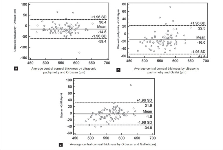
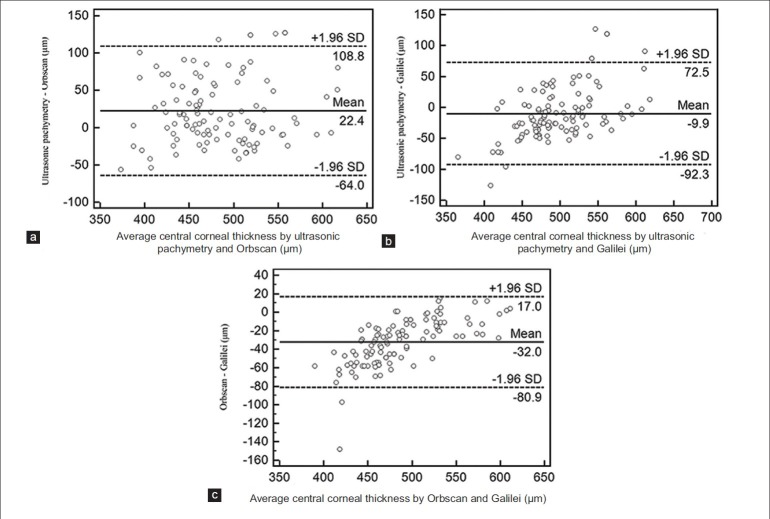
Methods: In this prospective study, CCT and PCT were measured in 88 eyes of 88 refractive surgery candidates and 128 eyes of 69 keratoconic patients with ultrasonic pachymetry, the Galilei, and Orbscan II. The readings by the three instruments were compared using one-way analysis of normal variance. Agreement among the three devices was assessed using Pearson and intraclass correlation coefficients, and Bland-Altman plots. The same analyses were employed to evaluate agreement between Galilei and Orbscan II for PCT measurement.
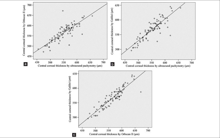
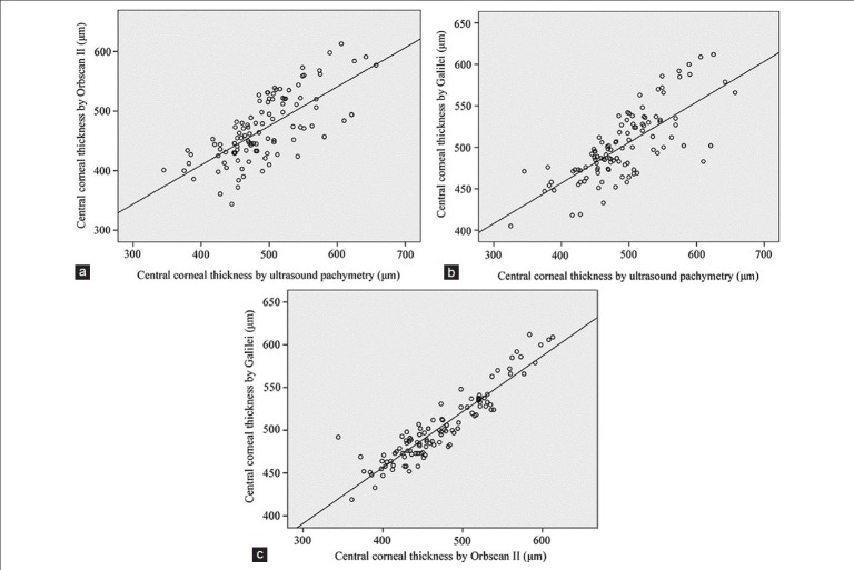
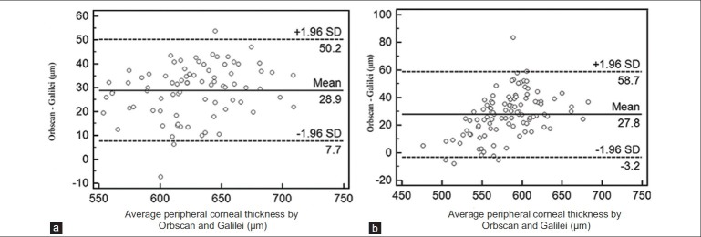
Results: In the normal group, mean CCT was 551.0±39.4, 566.9±33.5, and 565.5±40.9 μm measured by ultrasonic pachymetry, the Galilei, and Orbscan II, respectively (P<0.001). The corresponding figures in the keratoconus group were 492.0±61.7, 502.0±42.1, and 470.6±56.9 μm, respectively (P<0.001). Mean PCT was 612.5±35.3 and 640.9±38.0 μm in the normal group (P<0.001) and 567.6±35.2 and 595.1±41.4 μm in the keratoconus group (P<0.001) by the Galilei and Orbscan II, respectively. CCT and PCT measurements obtained by different devices were significantly correlated in both groups.
Conclusion: To measure CCT, the Galilei and Orbscan II can be used interchangeably in normal eyes, but not in keratoconic eyes. For PCT, there is a systematic error between measurements obtained by the Galilei and Orbscan II. However, it is possible to change optical pachymeter readings into those obtained by ultrasonic pachymetry using a constant.
Keywords: Central Corneal Thickness; Galilei; Keratoconus; Orbscan II; Peripheral Corneal Thickness; Ultrasonic Pachymetry.
Conflict of interest statement
Figures

References
-
- Rabinowitz YS, Rasheed K, Yang H, Elashoff J. Accuracy of ultrasonic pachymetry and videokeratography in detecting keratoconus. J Cataract Refract Surg. 1998;24:196–201. - PubMed
-
- Colin J, Cochener B, Savary G, Malet F. Correcting keratoconus with intracorneal rings. J Cataract Refract Surg. 2000;26:1117–1122. - PubMed
-
- Giasson C, Forthomme D. Comparison of central corneal thickness measurements between optical and ultrasound pachometers. Optom Vis Sci. 1992;69:236–241. - PubMed
-
- Rainer G, Petternel V, Findl O, Schmetterer L, Skorpik C, Luksch A, et al. Comparison of ultrasound pachymetry and partial coherence interferometry in the measurement of central corneal thickness. J Cataract Refract Surg. 2002;28:2142–2145. - PubMed
LinkOut - more resources
Full Text Sources