

Diabetes and retinal vascular dysfunction
- PMID: 25667739
- PMCID: PMC4307665
- DOI: 10.4103/2008-322X.143378
Diabetes and retinal vascular dysfunction
Abstract
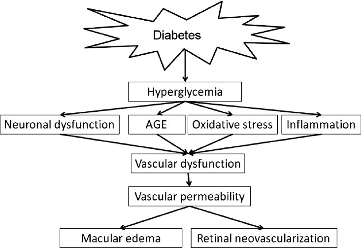
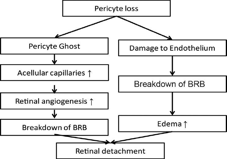
Diabetes predominantly affects the microvascular circulation of the retina resulting in a range of structural changes unique to this tissue. These changes ultimately lead to altered permeability, hyperproliferation of endothelial cells and edema, and abnormal vascularization of the retina with resulting loss of vision. Enhanced production of inflammatory mediators and oxidative stress are primary insults with significant contribution to the pathogenesis of diabetic retinopathy (DR). We have determined the identity of the retinal vascular cells affected by hyperglycemia, and have delineated the cell autonomous impact of high glucose on function of these cells. We discuss some of the high glucose specific changes in retinal vascular cells and their contribution to retinal vascular dysfunction. This knowledge provides novel insight into the molecular and cellular defects contributing to the development and progression of diabetic retinopathy, and will aid in the development of innovative, as well as target specific therapeutic approaches for prevention and treatment of DR.
Keywords: Diabetes; Inflammation; Oxidative Stress; Retinal vasculature; Thrombospondins.
Conflict of interest statement
Figures
References
-
- Runkle EA, Antonetti DA. The blood-retinal barrier: Structure and functional significance. Methods Mol Biol. 2011;686:133–148. - PubMed
-
- Kaur C, Foulds WS, Ling EA. Blood-retinal barrier in hypoxic ischaemic conditions: Basic concepts, clinical features and management. Prog Retin Eye Res. 2008;27:622–647. - PubMed
-
- Hosoya K, Tachikawa M. Inner blood-retinal barrier transporters: Role of retinal drug delivery. Pharm Res. 2009;26:2055–2065. - PubMed
-
- Klaassen I, Van Noorden CJ, Schlingemann RO. Molecular basis of the inner blood-retinal barrier and its breakdown in diabetic macular edema and other pathological conditions. Prog Retin Eye Res. 2013;34:19–48. - PubMed
-
- Antonetti DA, Barber AJ, Bronson SK, Freeman WM, Gardner TW, Jefferson LS, et al. Diabetic retinopathy: Seeing beyond glucose-induced microvascular disease. Diabetes. 2006;55:2401–2411. - PubMed
Publication types
Grants and funding
LinkOut - more resources
Full Text Sources
Other Literature Sources