Review
. 2015:77:179-200.
doi: 10.1146/annurev-physiol-021113-170255.

Cellular homeostasis and repair in the mammalian liver

Affiliations
Review

Cellular homeostasis and repair in the mammalian liver

Ben Z Stanger. Annu Rev Physiol. 2015.

Abstract

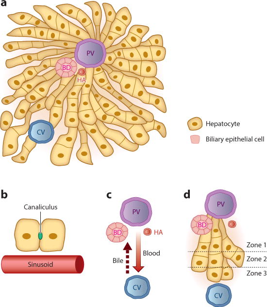
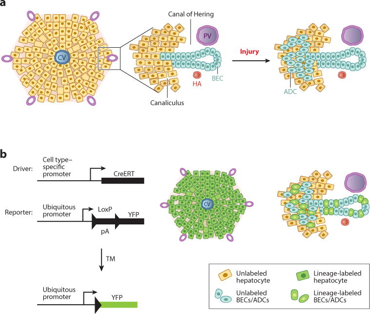
The mammalian liver is one of the most regenerative tissues in the body, capable of fully recovering mass and function after a variety of injuries. This factor alone makes the liver unusual among mammalian tissues, but even more atypical is the widely held notion that the method of repair depends on the manner of injury. Specifically, the liver is believed to regenerate via replication of existing cells under certain conditions and via differentiation from specialized cells--so-called facultative stem cells--under others. Nevertheless, despite the liver's dramatic and unique regenerative response, the cellular and molecular features of liver homeostasis and regeneration are only now starting to come into relief. This review provides an overview of normal liver function and development and focuses on the evidence for and against various models of liver homeostasis and regeneration.

Keywords: cellular reprogramming; homeostasis; injury; liver regeneration.


Figures

Figure 1
Figure 1
The liver lobule. (a) The liver is composed largely of two endoderm-derived cell types—hepatocytes and biliary epithelial cells—that are organized into functional units known as lobules. Blood enters the lobule through branches of the PV and HA and exits through branches of the CV to be returned to the systemic circulation. Bile makes its way to the BD, which in turn drains into the intestine to aid in digestion. (b) Blood passes through the lobule in specialized vascular channels (sinusoids). Bile is handled separately and is secreted into thin channels (canaliculi) between hepatocytes that carry the bile through the lobule and into the BD. (c) There is countercurrent flow of blood and bile: Blood flows in a portal-to-central direction, whereas bile flows in a central-to-portal direction. (d) The lobule is divided into zones, with hepatocytes closest to the PV composing zone 1 and those closest to the CV composing zone 3. Hepatocytes in different zones have different sets of functions. Abbreviations: BD, bile duct; CV, central vein; HA, hepatic artery; PV, portal vein.
Figure 2
Figure 2
Use of lineage tracing to study cellular dynamics in liver regeneration. (a) (Left) Schematic view of a liver lobule, with a CV in the center and the portal tracts at the periphery. (Middle) An enlargement of a portal tract reveals the canal of Hering: a transitional zone between the canaliculi and the bile ducts that is asymmetrically lined by hepatocytes and BECs. (Right) Upon injury, ADCs (oval cells) emerge near the portal tracts. (b) In vivo Cre-based lineage tracing requires the use of a Cre-expressing genetic driver, which provides a means of marking cells in a specific manner, and a reporter, which allows for the detection of cells that have been marked as a result of Cre activity. In one iteration, a TM-inducible Cre (CreERT) becomes active only in the presence of the synthetic estrogen analog TM, allowing for labeling of cells (green) that have active CreERT transcription from the cell type–specific promoter employed. On the right are examples of the types of labeling that could be envisioned with a driver that permits labeling of hepatocytes with high efficiency (key, top) or with one that permits labeling of ADCs and BECs with medium efficiency (key, bottom). Abbreviations: ADC, atypical ductal cell; BEC, biliary epithelial cell; CV, central vein; HA, hepatic artery; pA, RNA polyadenylation signal; PV, portal vein; TM, tamoxifen; YFP, yellow fluorescent protein.

References

    1. Slack JM. Origin of stem cells in organogenesis. Science. 2008;322:1498–501. - PubMed
    1. Weissman IL. Stem cells: units of development, units of regeneration, and units in evolution. Cell. 2000;100:157–68. - PubMed
    1. Simon A, Tanaka EM. Limb regeneration. Wiley Interdiscip Rev Dev Biol. 2013;2:291–300. - PubMed
    1. Tornini VA, Poss KD. Keeping at arm’s length during regeneration. Dev Cell. 2014;29:139–45. - PMC - PubMed
    1. Thorel F, Nepote V, Avril I, Kohno K, Desgraz R, et al. Conversion of adult pancreatic α-cells to β-cells after extreme β-cell loss. Nature. 2010;464:1149–54. - PMC - PubMed

Publication types