. 2015 Apr 1;284(1):65-78.
doi: 10.1016/j.taap.2015.01.022. Epub 2015 Feb 8.

Motoneuron axon pathfinding errors in zebrafish: differential effects related to concentration and timing of nicotine exposure

Affiliations

Motoneuron axon pathfinding errors in zebrafish: differential effects related to concentration and timing of nicotine exposure

Evdokia Menelaou et al. Toxicol Appl Pharmacol. .

Abstract

Nicotine exposure during embryonic stages of development can affect many neurodevelopmental processes. In the developing zebrafish, exposure to nicotine was reported to cause axonal pathfinding errors in the later born secondary motoneurons (SMNs). These alterations in SMN axon morphology coincided with muscle degeneration at high nicotine concentrations (15-30 μM). Previous work showed that the paralytic mutant zebrafish known as sofa potato exhibited nicotine-induced effects onto SMN axons at these high concentrations but in the absence of any muscle deficits, indicating that pathfinding errors could occur independent of muscle effects. In this study, we used varying concentrations of nicotine at different developmental windows of exposure to specifically isolate its effects onto subpopulations of motoneuron axons. We found that nicotine exposure can affect SMN axon morphology in a dose-dependent manner. At low concentrations of nicotine, SMN axons exhibited pathfinding errors, in the absence of any nicotine-induced muscle abnormalities. Moreover, the nicotine exposure paradigms used affected the 3 subpopulations of SMN axons differently, but the dorsal projecting SMN axons were primarily affected. We then identified morphologically distinct pathfinding errors that best described the nicotine-induced effects on dorsal projecting SMN axons. To test whether SMN pathfinding was potentially influenced by alterations in the early born primary motoneuron (PMN), we performed dual labeling studies, where both PMN and SMN axons were simultaneously labeled with antibodies. We show that only a subset of the SMN axon pathfinding errors coincided with abnormal PMN axonal targeting in nicotine-exposed zebrafish. We conclude that nicotine exposure can exert differential effects depending on the levels of nicotine and developmental exposure window.

Keywords: Axonal pathfinding; Dual labeling; Embryonic spinal cord; Muscle degeneration.


Figures

Figure 1
Figure 1. Secondary motoneuron axons and the effects of nicotine exposure on their axonal pathfinding
A) Photomicrograph of a 72-hpf larval Tg(isl1:GFP) zebrafish immunolabeled with the zn5 antibody (red) which specifically labels the main nerve of fasciculated SMNs axons. This zebrafish was exposed to nicotine (30µM) embryonically (22–72 hpf) and exhibits axonal pathfinding errors in 4 consecutive segments. B) zn5 immunostaining is shown in the Tg(isl1:GFP) larval zebrafish that expresses GFP (white) in a subset of secondary motoneurons (SMN). Zn5-positive axons colocalize with GFP-positive dorsal projecting SMN axons. C) Left segment shows a lateral view of different subpopulations of secondary motoneurons (SMNs, shown in orange, red and blue) located in ventral spinal cord (marked by dotted lines). Axons exit at mid-segmental root and extend dorsally (D, red), ventrally (V, blue), and mediolaterally (ML, orange). Right segment illustrates normal pathfinding of SMN axons as visualized by zn5 labeling. D) Examples of abnormal axonal pathfinding are shown in each segment in the dorsal and ventral myotome. Note that abnormal phenotypes can be seen for dorsal, ventral, and mediolateral projecting axons. Horizontal myoseptum: hm. Illustration is not drawn to scale. Scale bar, 20 µm.
Figure 2
Figure 2. Nicotine-induced effects on SMN axons depend upon nicotine concentration and window of exposure
Ai) Zn5 immunoreactivity reveals SMN axons and their somata in 72-hpf control larva and corresponding zebrafish larvae exposed to varying concentrations of nicotine, 1µM (Bi), 5µM (Ci), 15µM (Di) and 30µM (Ei) from 22–72 hpf. Note the abnormal motoneuron axon trajectories in the nicotine-exposed zebrafish when compared to their control counterparts. Fi) At 72 hpf, the number of hemisegments with motoneuron axons that projected normally in the dorsal, ventral, and mediolateral myotomes over the total number of hemisegments analyzed in each fish was quantified and expressed as a percentage. Zebrafish exposed to 1µM (516 segments; 18 fish), 5µM (355 segments; 14 fish), 15µM (436 segments; 16 fish), and 30µM nicotine (359 segments; 14 fish) exhibited more hemisegments with abnormal motoneuron axon morphologies when compared to their stage-matched controls (455 segments; 17 fish). Asterisks in Fi denote statistical significance, p value < 0.001. Nicotine groups compared against the control using a post hoc Holm-Sidak test. Aii–Eii) Same as in Ai–Ei but for nicotine exposure window between 12–30 hpf. Fii) Quantification of nicotine-induced effects on SMN axons for exposure window 12–30 hpf. Zebrafish exposed to 1µM (176 segments; 7 fish) and 5µM nicotine (139 segments; 6 fish) were not significantly affected whereas when exposed at 15µM (125 segments; 5 fish) and 30µM (146 segments; 6 fish) were affected when compared to stage-matched controls (152 segments; 6 fish). Filled arrows indicate pathfinding errors in dorsal projecting axons. Open arrows indicate pathfinding errors in ventral projecting axons. Arrowheads indicate errors in ventromedial projecting axons. Quantification shown in Fi and Fii is from a single representative experiment for each nicotine exposure paradigm (also see, Supplemental Fig.1). Asterisks in Fii denote statistical significance, p value < 0.05. Nicotine groups were compared against the control using a post hoc Dunn’s test. Scale bars, 20 µm.
Figure 3
Figure 3. Morphology of slow muscle fibers following nicotine exposure
F59 immunoreactivity was used to reveal slow muscle fibers in 72-hpf zebrafish larvae exposed to varying concentrations of nicotine. Representative photomicrographs reveal multiple slow muscle fibers in the field of view from control larvae (Ai) and corresponding zebrafish larvae exposed to 1µM (Bi) 30µM (Ci) nicotine from 22–72 hpf. Di) Quantification of slow muscle fiber widths in 72 hpf zebrafish for exposure window between 22–72 hpf (control: 268 muscle fibers, 7 fish; 1µM: 237 muscle fibers, 7 fish; 5µM: 165 muscle fibers, 5 fish; 15µM: 207 muscle fibers, 7 fish; 30µM: 198 muscle fibers, 7 fish). Aii–Cii) Same as in Ai–Ci but for nicotine exposure window between 12–30 hpf. Dii) Quantification of slow muscle fiber widths in 72-hpf zebrafish for exposure window between 12–30 hpf (control: 363 muscle fibers, 12 fish; 1µM: 256 muscle fibers, 10 fish; 5µM: 463 muscle fibers, 16 fish; 15µM: 398 muscle fibers, 13 fish; 30µM: 204 muscle fibers, 9 fish). Asterisk denotes statistical significance, p value < 0.05. Nicotine groups were compared against the control using a post hoc Holm-Sidak test. Scale bars, 20 µm.
Figure 4
Figure 4. Effect of embryonic nicotine exposure on dorsal projecting SMN axons
A) Drawings depict a normal SMN axon projecting dorsally (grey) superimposed with observed nicotine-induced abnormal phenotypes (black). B) Dorsal projecting axons in 72-hpf zebrafish with abnormal phenotypes following a nicotine exposure between 12–30 hpf and 22–72 hpf. Dotted line denotes the boundaries of the spinal cord. The number of fish and hemisegments analyzed for each nicotine exposure paradigm is the same as in Table 1. Asterisk denotes statistical significance, p value < 0.05; double asterisks, p value < 0.001. Nicotine groups were compared against the control using a post hoc Holm-Sidak test. Cartoon is not drawn to scale.
Figure 5
Figure 5. Three phenotypically distinct errors emerge on dorsal projecting SMN axons following embryonic nicotine exposure
A) Left, low magnification photomicrograph (zn5 immunoreactivity) reveals normal SMN axon trajectories (scale bar, 50 µm). Middle, diagram illustrates the normal trajectories of dorsal, ventral, and mediolateral motoneuron axons and the box outlines the region shown to the right. Right, high magnification photomicrograph reveals the normal trajectory path (arrowheads) of the dorsal projecting axon. Nicotine-induced phenotypes of dorsal projecting axons were categorized as “trajectory” (B), “split” (C) and “stall” (D) axonal errors. For each category, 4 representative example phenotypes are shown from different nicotine exposed fish. B) “Trajectory” error refers to dorsal motoneuron axons that reach their dorsal most target but they drift (arrowheads) past their turning point and make sharp turns as they traject (open arrows). C) As the main nerve bundle of dorsal projecting SMN axons projects in the periphery, it splits (filled arrows) mainly into two different nerve fibers. D) Motor axons completely stall (dotted circle) as they exit the ventral root and fail to reach their dorsal most targets in the periphery. Arrowheads in D denote zn5-positive axons that have started to target dorsally after being stalled at the exit point. Scale bars, 20 µm.
Figure 6
Figure 6. The nicotine-induced phenotypes of dorsal projecting SMN axons depend on the length of the exposure and nicotine concentration
A–C) Left, example cartoons depict one dorsal projecting SMN axons that has a normal projection and two abnormal phenotypes. Right, Quantification of the dorsal projecting SMN axons that have the corresponding abnormal phenotypes shown to the left (“trajectory” error, A; “split” axon error, B; “stall” axon error, C). Normal SMN axon phenotypes are shown in grey and abnormal phenotypes are shown in black. Dotted line denotes the boundaries of spinal cord. The number of fish and hemisegments analyzed for each nicotine exposure paradigm is the same as in Table 1. Dorsal is to the top and rostral is to the left. Asterisk denotes statistical significance, p value < 0.05. Nicotine exposure groups were compared against the control using a post hoc Holm-Sidak (A) or Dunn’s test (B and C). Cartoons are not drawn to scale.
Figure 7
Figure 7. anti-chrn2b labels a subset of primary motoneuron somata and axons in zebrafish embryos
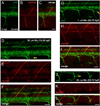
A) Left, image of CaP motoneuron detected by anti-chrn2b in a 28-hpf embryo. The white open and white arrows indicate the soma and axon of the putative CaP motoneuron, respectively (same in B). B) Image of CaP motoneuron axon and soma as well as a dorsal projecting axon (yellow arrow) detected by anti-chrn2b in a 30 hpf embryo. C) Image of 32-hpf embryo labeled with anti-chrn2b. Yellow arrow denotes the putative MiP motoneuron axon. White open arrow points to CaP motoneuron soma. Yellow circle highlights the presumptive soma of MiP motoneuron. D) Image from a 28-hpf embryo focusing on three ventral mytomes labeled with anti-chrn2b. E) Image from same fish in panel D now showing anti-chrn2b coupled with znp-1 labeling. F) Dual immunolabeling in a 31-hpf embryo using anti-chrn2b and the zn5 antibody. The anti-chrn2b detected the ventral and dorsal projecting axons as shown from 1 body segment. The zn5 antibody labeled SMN somata and ventral projecting SMN axons. Note that the two antibodies detect two non-overlapping axons ventrally and dorsally only the earlier born MiP motoneuron axon (yellow arrow) projects into the periphery. G) Image of a dorsal projecting motoneuron axon labeled with znp-1 in a 33-hpf embryo. H) Image is from the same segment shown in panel G. The MiP axon was detected by anti-chrn2b (yellow arrow). I) Merging of panels G and H reveals colocalization of znp-1 and anti-chrn2b signal at the level of the dorsal projecting axon. J) Image of a 24-hpf, pargmn2Et embryo. White arrows denote putative MiP motoneuron axons. K) Image of a 34-hpf pargmn2Et embryo. White arrow denotes MiP axon. L) Image is from the same segment shown in panel K where the embryo was labeled with anti-chrn2b. Yellow arrow denotes dorsal projecting axon. M) Merging of panels K and L reveals co-localization of GFP signal and anti-chrn2b axonal signal at the level of the MiP motoneuron axon. See Supplemental Table 1 for number of fish analyzed for each developmental time point. Scale bars; 10 µm.
Figure 8
Figure 8. Simultaneous examination of nicotine-induced effects onto primary and secondary motoneuron axons
A–C) Representative photomicrographs from a 72-hpf control larva labeled with the zn5 antibody and anti-chrn2b. Photomicrographs from 3 different 72-hpf larvae labeled with the zn5 antibody and anti-chrn2b and exposed to either 30µM nicotine from 12–30 hpf (D–F), 1µM nicotine from 22–72 hpf (G–I) or 30µM nicotine from 22–72 hpf (J–L). Yellow arrows in each exposure example denote SMN axons that extended too far caudally before turning dorsally. See Supplemental Table 1 for number of fish analyzed for each nicotine exposure paradigm. Scale bars; 20 µm.
Figure 9
Figure 9. The nicotine-induced “stall” and “duplication” errors are specific to SMN axons
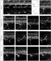
A–C) Photomicrographs of a 72-hpf larva exposed to 30µM nicotine from 22–72 hpf. The zn5-positive SMN dorsal projecting axons (yellow arrows in A) have not extended into the periphery and appear to have stalled. B–C) Anti-chrn2b antibody detects MiP motoneuron axons in the periphery whereas zn5-positive SMN axons have not extended into the periphery. D) Image of a 72-hpf larva exposed to 30µM nicotine from 22–72 hpf. There is a duplicated SMN nerve root shown by the yellow arrow. E) Photomicrograph of the fish shown in D labeled with anti-chrn2b. The anti-chrn2b-positive dorsal and ventral projecting axons extended out into the periphery in these 3 consecutive segments. There was no ventral nerve root duplication that matched the zn5-positive one in panel D (yellow arrow). See Supplemental Table 1 for number of fish analyzed for each nicotine exposure paradigm. Scale bars; 20 µm.
Figure 10
Figure 10. Differential effects along the motoneuron axon path following nicotine exposures
A–C) Photomicrographs of a 72-hpf larva labeled with zn5 and anti-chrn2b capture motoneuron axon extension within 1 segment just below spinal cord (ventral edge of spinal cord indicated by white arrowheads). This larva was exposed to 30µM from 22–72 hpf. Note that the zn5-positive axon appears stalled at this magnification whereas the anti-chrn2b-positive axon (white arrow in panel B) takes a dorsal trajectory. Yellow arrow in A points to a weak zn5 signal; likely a few axons which have migrated dorsally. The asterisk in A provides a reference point of the magnified area in D–F. D–F) The images in A–C shown at higher magnification. Zn5-positive axons appear to migrate to the periphery using the anti-chrn2b - positive axon as a guide. Conventions are the same as in A–C. G–I) Photomicrographs of a 72-hpf larva labeled with zn5 and anti-chrn2b. This larva was exposed to 30µM from 22–72 hpf. Zn5-positive axons appear to use the anti-chrn2b-positive axon as a guide when navigating below spinal cord (marked by asterisks). Once in the distal dorsal periphery, the zn5-positive axons exhibit pathfinding errors (yellow arrow) whereas the anti-chrn2b-positive axon targeted the periphery using a slightly different dorsal path (white arrow). The zebrafish larva presented under panels A–F was presented earlier in Fig. 2Ei. See Supplemental Table 1 for number of fish analyzed for each nicotine exposure paradigm. Scale bars; 10 µm in C and F, 20 µm in I.

Similar articles

Cited by

References

    1. Ackerman KM, Nakkula R, Zirger JM, Beattie CE, Boyd RT. Cloning and spatiotemporal expression of zebrafish neuronal nicotinic acetylcholine receptor alpha 6 and alpha 4 subunit RNAs. Dev Dyn. 2009;238(4):980–992. - PMC - PubMed
    1. An MC, Lin W, Yang J, Dominguez B, Padgett D, Sugiura Y, Aryal P, Gould TW, Oppenheim RW, Hester ME, Kaspar BK, Ko C, Lee K. Acetylcholine negatively regulates development of the neuromuscular junction through distinct cellular mechanisms. PNAS. 2010;107(23):10702–10707. - PMC - PubMed
    1. Balciunas D, Davidson AE, Sivasubbu S, Hermanson SB, Welle Z, Ekker SC. Enhancer trapping in zebrafish using the Sleeping Beauty transposon. BMC Genomics. 2004;5:62. - PMC - PubMed
    1. Beattie CE, Melancon E, Eisen JS. Mutations in the stumpy gene reveal intermediate targets for zebrafish motor axons. Development. 2000;127(12):2653–2662. - PubMed
    1. Behra M, Cousin X, Bertrand C, Vonesch JL, Biellmann D, Chatonnet A, et al. Acetylcholinesterase is required for neuronal and muscular development in the zebrafish embryo. Nature Neuroscience. 2002;5(2):111–118. - PubMed

Publication types

MeSH terms

LinkOut - more resources