. 2015 Mar 1:569:32-44.
doi: 10.1016/j.abb.2015.02.002. Epub 2015 Feb 7.

The invertebrate Caenorhabditis elegans biosynthesizes ascorbate

Affiliations

The invertebrate Caenorhabditis elegans biosynthesizes ascorbate

Alexander N Patananan et al. Arch Biochem Biophys. .

Abstract

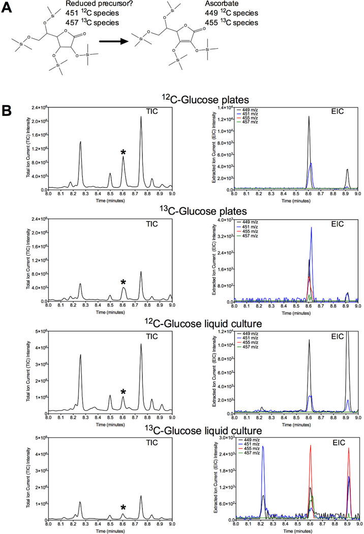
l-Ascorbate, commonly known as vitamin C, serves as an antioxidant and cofactor essential for many biological processes. Distinct ascorbate biosynthetic pathways have been established for animals and plants, but little is known about the presence or synthesis of this molecule in invertebrate species. We have investigated ascorbate metabolism in the nematode Caenorhabditis elegans, where this molecule would be expected to play roles in oxidative stress resistance and as cofactor in collagen and neurotransmitter synthesis. Using high-performance liquid chromatography and gas-chromatography mass spectrometry, we determined that ascorbate is present at low amounts in the egg stage, L1 larvae, and mixed animal populations, with the egg stage containing the highest concentrations. Incubating C. elegans with precursor molecules necessary for ascorbate synthesis in plants and animals did not significantly alter ascorbate levels. Furthermore, bioinformatic analyses did not support the presence in C. elegans of either the plant or the animal biosynthetic pathway. However, we observed the complete (13)C-labeling of ascorbate when C. elegans was grown with (13)C-labeled Escherichia coli as a food source. These results support the hypothesis that ascorbate biosynthesis in invertebrates may proceed by a novel pathway and lay the foundation for a broader understanding of its biological role.

Keywords: Ascorbic acid; Biosynthetic pathway; Caenorhabditis elegans; Invertebrates; Vitamin C.


Figures

Figure 1
Figure 1. HPLC detection of ascorbate in extracts of mixed populations of C. elegans
(A) Ascorbate standard separated by reversed-phase HPLC. The black line represents 3.9 µg of ascorbic acid dissolved in a final concentration of 1.7% metaphosphoric acid, 1.7 mM EDTA, 1.7 mM tris(2-carboxyethyl) phosphine hydrochloride, and 130 mM sodium citrate, pH 8, and separated on a reversed-phase HPLC column as described in the “Materials and Methods” section. 2 units of ascorbate oxidase (AAO) were added to the standard to confirm the elution of ascorbate as described in “Materials and Methods” (red line). (B) Reversed-phase HPLC analysis of a C. elegans extract prepared from a mixed population of animals obtained by washing two NGM plates (~142,000 worms) as described in the “Materials and Methods.” 55 µl of sample was injected onto the HPLC (black line). 2 units of AAO was added to confirm the elution of ascorbate (red line).
Figure 2
Figure 2. GC-MS detection of ascorbate in mixed populations of C. elegans
(A) GC-MS fragmentation pattern of ascorbate derivatized with BSTFA/TMCS from the NIST Mass Spectral Library 08. An enlarged view of the m/z for the specific fragments labeled a–g are shown in the panels at the top. In the structures represented below the panel, the fragment of ascorbate is shown in red; when multiple fragments are possible for a particular m/z, the alternative fragment is shown in blue. Among the peaks, the 332 m/z fragment represents one of the most intense signals. (B). GC-MS analysis of a BSTFA/TMCS derivatized extract of mixed population of C. elegans obtained from ten NGM plates was performed as described in the “Materials and Methods.” The total ion current (TIC) and 332 m/z extracted ion current (97) are represented by the black and red traces, respectively. The asterisk (*) denotes the ascorbate peak. (C) The MS fragmentation pattern of the species denoted by the asterisk identified in panel B. (D) GC-MS analysis of a standard of 150 ng of ascorbate derivatized in BSTFA/TMCS to confirm the elution time of ascorbate as described in the “Materials and Methods.” The TIC and 332 m/z EIC are represented by the black and red traces, respectively. The asterisk (*) denotes the ascorbate peak. The peak located at 7.4 min is an artifact identified as derivatized triethylene glycol.
Figure 3
Figure 3. Lack of detectable ascorbate in C. elegans growth media and extraction reagent
(A) TIC and (B) 332 m/z EIC were analyzed by GC-MS of an extract derived from a mixed population of C. elegans, 0.5 mM BHT in methanol extraction buffer, S medium, M9 medium, sucrose, E. coli extract, and LB medium. Samples were derivatized with BSTFA/TMCS as described in the “Materials and Methods.” The dashed red line denotes the elution position of ascorbate. Although small peaks were found in the TIC and 332 m/z EIC of sucrose and E. coli, further analysis of the mass spectrum at these elution times for the sucrose (C) and E. coli (D) samples reveal that the fragmentation patterns do not match ascorbate, but rather weakly match derivatized ribofuranose and phosphate, respectively.
Figure 4
Figure 4. C. elegans deficient in mcp-1 have ascorbate levels equivalent to wild type
(A) mcp-1 (tm2679) extracts were prepared from two NGM plates containing a mixed population of worms (approximately 208,000 animals) and analyzed by reversed-phase HPLC as described in the “Materials and Methods” (black line). The extracts were treated with 2 units of AAO to confirm the existence of ascorbate (red line). (B) Ascorbate levels were quantified by HPLC and compared between the N2 wild type and mcp-1 (tm2679) extracts prepared from two NGM plates containing a mixed population of worms (N2, 43,700–142,400 animals; mcp-1 (tm2679), 106,000–318,500) as described in the “Materials and Methods.” The points represent three independent samples, the horizontal line the average, and the error bars represent standard deviations. Statistical significance was determined using an unpaired, two-tailed Student’s t-test. (C) N2 and mcp-1 (tm2679) extracts were prepared in 0.5 mM BHT in methanol and analyzed by GC-MS as described in the “Materials and Methods.” The area of the ascorbate 332 m/z EIC peak in each sample was determined and compared against the area of the serine 204 m/z EIC peak. The points represent four independent samples, the horizontal line is the average, and the error bars represent standard deviations. Statistical significance was determined using an unpaired, two-tailed Student’s t-test.
Figure 5
Figure 5. Ascorbate levels are highest in the egg stage of C. elegans
Extracts of eggs, L1 larvae, and mixed populations derived from 9–83, 5–69, and 38–455 mg wet weight of animals, respectively, were prepared as described in the “Materials and Methods” section. Ascorbate was separated by reversed-phase HPLC and quantified. Five, sixteen, and twelve independent samples were analyzed for the eggs, L1 larvae, and mixed population of worms, respectively. The data points represent independent samples, the horizontal line is the average, and the error bars represent standard deviations. Statistical significance was determined using an unpaired, two-tailed Student’s t-test.
Figure 6
Figure 6. The addition of ascorbate precursors from known ascorbate biosynthetic pathways does not increase ascorbate levels in C. elegans
(A–C) C. elegans membrane, cytosolic, and intact worm fractions were incubated with a final concentration of 6.7 mM L-gulono-1,4-lactone for 2 h at room temperature and analyzed by HPLC as described in the “Materials and Methods.” Asterisks (*) denote the elution position of ascorbate. (D) L1 larvae obtained from 80 NGM plates were incubated in a final concentration of 1.7 mM D-galactose, L-gulono-1,4-lactone, D-galacturonic acid, D-glucose, or D-glucuronic acid for 2 h at 20 °C and 160 rpm, before the samples were lysed and analyzed by reversed-phase HPLC according to the “Materials and Methods” section. The variability in ascorbate elution between panels A–C and D is due to a change in HPLC column.
Figure 7
Figure 7. Paraquat and ethanol stress do not increase ascorbate levels in C. elegans
(A) A mixed population of animals was obtained from approximately six NGM plates and incubated in S-medium supplemented with OP50 E. coli and 0, 50, 100, and 200 µM paraquat for 7 d as described in the “Materials and Methods.” GC-MS analysis was performed as described in the “Materials and Methods” and the amount of ascorbate in samples was analyzed by comparing the EIC area of ascorbate (332 m/z) to that of serine (204 m/z). The points represent four independent samples, the horizontal line is the average, and the error bars denote standard deviations. Statistical significance was determined using an unpaired, two-tailed Student’s t-test. (B) Mixed populations of animals from 80 NGM plates were incubated in a microcentrifuge tube at 20 °C and 160 rpm in M9 medium supplemented with 0, 1%, 5%, or 10% ethanol. After incubation for 39 h, the animals were pelleted, washed, lysed, and analyzed by reversed-phase HPLC according to the “Materials and Methods” section. The data points represent two independent samples and the horizontal line denotes the average. Statistical significance was determined using an unpaired, two-tailed Student’s t-test.
Figure 8
Figure 8. Incorporation of 13C into C. elegans ascorbate from 13C-labeled E. coli food
(A) Expected mass change in ascorbate and the possible reduced precursor species due to uniform13C labeling. (B) A mixed population of animals were incubated with 12C6-D-glucose or 13C6-D-glucose-labeled E. coli on plates or in liquid culture at 20 °C for 85 h as described in the “Materials and Methods” section and analyzed by GC-MS. The asterisk denotes the position of ascorbate.
Figure 9
Figure 9. Ascorbate fragmentation pattern from C. elegans fed unlabeled or 13C-labeled E. coli
Mixed populations of animals were incubated with unlabeled OP50 E. coli or OP50 E. coli labeled with 13C6-D-glucose on (A) NGM plates or (B) in liquid culture for 85 h at 20 °C and analyzed by GC-MS as described in Figure 8 and the “Materials and Methods.” Ascorbate fragmentation data for animals incubated with unlabeled or 13C-labeled OP50 E. coli is shown in blue and red, respectively, and overlaid for direct comparison. The peaks labeled a, b, c, d, e, f, and g correspond to major ascorbate fragmentation peaks shown in Figure 2 and include the 117, 147, 205, 259, 304, 332, and 449 m/z ions. A magnified view of these fragmentation peaks is shown in the inset figures in panels A and B, with the y-axis representing relatively intensity and the x-axis denoting the m/z.

Similar articles

Cited by

References

    1. Du J, Cullen JJ, Buettner GR. Ascorbic acid: chemistry, biology and the treatment of cancer. Biochim. Biophys. Acta. 2012;1826:443–457. - PMC - PubMed
    1. Buettner GR, Jurkiewicz BA. Catalytic metals, ascorbate and free radicals: combinations to avoid. Radiat. Res. 1996;145:532–541. - PubMed
    1. Linster CL, Clarke SG. L-ascorbate biosynthesis in higher plants: the role of VTC2. Trends Plant Sci. 2008;13:567–573. - PMC - PubMed
    1. Gao Q, Zhang L. Ultraviolet-B-induced oxidative stress and antioxidant defense system responses in ascorbate-deficient vtc1 mutants of Arabidopsis thaliana . J. Plant Physiol. 2008;165:138–148. - PubMed
    1. Barth C, De Tullio M, Conklin PL. The role of ascorbic acid in the control of flowering time and the onset of senescence. J. Exp. Bot. 2006;57:1657–1665. - PubMed

Publication types

MeSH terms