. 2015 Sep:114:87-99.
doi: 10.1016/j.visres.2015.01.016. Epub 2015 Feb 7.

Characteristics of fixational eye movements in amblyopia: Limitations on fixation stability and acuity?

Affiliations

Characteristics of fixational eye movements in amblyopia: Limitations on fixation stability and acuity?

Susana T L Chung et al. Vision Res. 2015 Sep.

Abstract

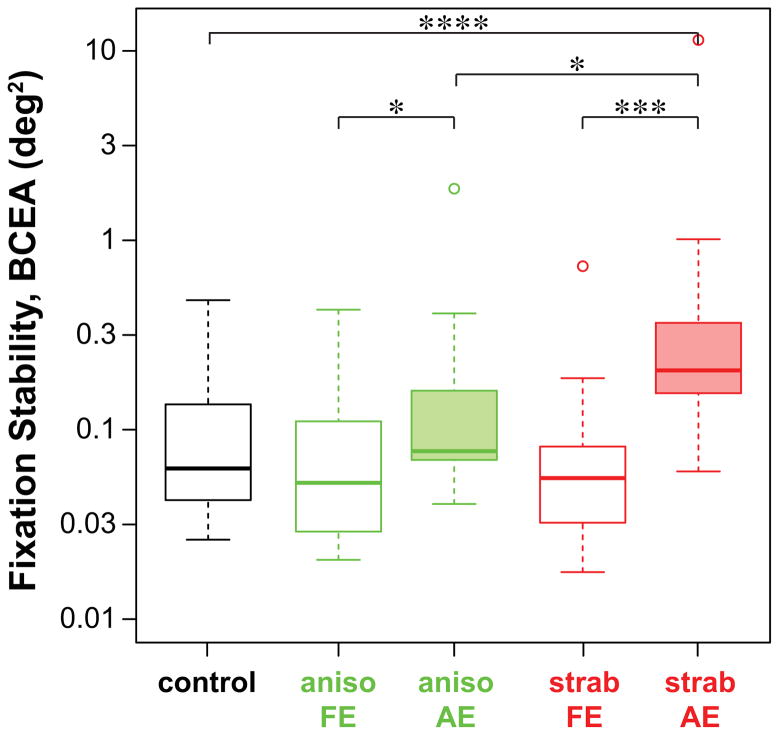
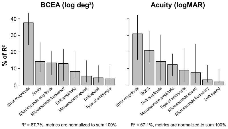
Persons with amblyopia, especially those with strabismus, are known to exhibit abnormal fixational eye movements. In this paper, we compared six characteristics of fixational eye movements among normal control eyes (n=16), the non-amblyopic fellow eyes and the amblyopic eyes of anisometropic (n=14) and strabismic amblyopes (n=14). These characteristics include the frequency, magnitude of landing errors, amplitude and speed of microsaccades, and the amplitude and speed of slow drifts. Fixational eye movements were recorded using retinal imaging while observers monocularly fixated a 1° cross. Eye position data were recovered using a cross-correlation procedure. We found that in general, the characteristics of fixational eye movements are not significantly different between the fellow eyes of amblyopes and controls, and that the strabismic amblyopic eyes are always different from the other groups. Next, we determined the primary factors that limit fixation stability and visual acuity in amblyopic eyes by examining the relative importance of the different oculomotor characteristics, adding acuity (for fixation stability) or fixation stability (for acuity), and the type of amblyopia, as predictive factors in a multiple linear regression model. We show for the first time that the error magnitude of microsaccades, acuity, amplitude and frequency of microsaccades are primary factors limiting fixation stability; while the error magnitude, fixation stability, amplitude of drifts and amplitude of microsaccades are the primary factors limiting acuity. A mediation analysis showed that the effects of error magnitude and amplitude of microsaccades on acuity could be explained, at least in part, by their effects on fixation stability.

Keywords: Amblyopia; Fixation stability; Fixational eye movements; Microsaccades; Slow drifts; Visual acuity.


Figures

Figure 1
Figure 1
Sample eye position traces of an amblyopic observer with anisometropia (top) and another with strabismus (bottom). Traces are shown for the non-amblyopic fellow eye (FE) and the amblyopic eye (AE). For clarity, the vertical (blue) and horizontal (red) eye position traces are offset vertically in each panel.
Figure 2
Figure 2
Box-and-whisker plots comparing the frequency of microsaccades, amplitude of microsaccades, speed of microsaccades, error magnitude of microsaccades, amplitude of slow drifts and speed of slow drifts for five groups of eyes: control eyes, non-amblyopic fellow eyes of anisometropic amblyopes (aniso FE), amblyopic eyes of anisometropic amblyopes (aniso AE), non-amblyopic fellow eyes of strabismic amblyopes (strab FE) and amblyopic eyes of strabismic amblyopes (strab AE). The upper and lower bound of each box represent the 75th and 25th percentiles of the distribution, and the median is represented by the thick line inside the box. The top and bottom ends of the whisker represent the 95th and 5th percentiles of the distribution, respectively. Outliers, if present, are represented by individual circles. The number of asterisks indicates the level of significance of the pairwise comparison according to the standard notation:* p ≤ 0.05, ** p ≤ 0.01, *** p ≤ 0.001, ***** p ≤ 0.0001.
Figure 3
Figure 3
Box-and-whisker plots comparing fixation stability, quantified as the bivariate contour ellipse area (BCEA, in deg2) for the five groups of eyes. Details of the boxes and whiskers are as in Figure 2.
Figure 4
Figure 4
Percentage of R2 of BCEA (log transformed values) and acuity (logMAR) accounted for by each of the parameter listed on the x-axes, as determined using the R package relaimpo. The relative importances of these parameters are ranked according to the lmg metric. Error bars represent ±95% confidence intervals estimated using bootstrappings, based on 10,000 resamplings.
Figure 5
Figure 5
Scatter plots of the relationship between each “important factor” that can predict either fixation stability or visual acuity. These factors are: fixation stability (log-transformed BCEA values were used here), visual acuity (logMAR), error magnitude (log-transformed), amplitude of microsaccades (log-transformed), amplitude of slow drifts (log-transformed) and frequency of microsaccades. The correlation coefficients for each pair of variables are given in the lower half of the figure. All correlation coefficients are significant at the p ≤ 0.05 level except for the pairs between frequency of microsaccades and acuity, error magnitude, amplitude of microsaccades, and amplitude of slow drifts (correlation coefficients shown in light gray).
Figure 6
Figure 6
A schematic figure illustrating the mediation model. The mediation model decomposes the total effect of an independent variable (in our case, error magnitude or amplitude of microsaccades) on the dependent variable (in our case, visual acuity) into two components: the direct effect (c) and the indirect effect through a mediator (in our case, fixation stability), quantified by a*b. The total effect is then represented by c+a*b. The ratio of the indirect effect to the total effect represents the proportion of the effect explained by the mediator.

Similar articles

Cited by

References

    1. Aytekin M, Victor JD, Rucci M. The visual input to the retina during natural head-free fixation. Journal of Neuroscience. 2014;34:12701–12715. - PMC - PubMed
    1. Bedell HE, Flom MC, Barbeito R. Spatial aberrations and acuity in strabismus and amblyopia. Investigative Ophthalmology & Visual Science. 1985;26:909–916. - PubMed
    1. Bedell HE, Yap YL, Flom MC. Fixational drift and nasal-temporal pursuit asymmetries in strabismic amblyopes. Investigative Ophthalmology & Visual Science. 1990;31:968–976. - PubMed
    1. Bland JM, Altman DG. Statistical methods for assessing agreement between two methods of clinical measurement. Lancet. 1986;1:307–310. - PubMed
    1. Bonneh YS, Sagi D, Polat U. Spatial and temporal crowding in amblyopia. Vision Research. 2007;47:1950–1962. - PubMed

Publication types