. 2015 May;35(5):861-72.
doi: 10.1038/jcbfm.2015.2. Epub 2015 Feb 11.

Intracranial pressure elevation reduces flow through collateral vessels and the penetrating arterioles they supply. A possible explanation for 'collateral failure' and infarct expansion after ischemic stroke

Affiliations

Intracranial pressure elevation reduces flow through collateral vessels and the penetrating arterioles they supply. A possible explanation for 'collateral failure' and infarct expansion after ischemic stroke

Daniel J Beard et al. J Cereb Blood Flow Metab. 2015 May.

Abstract

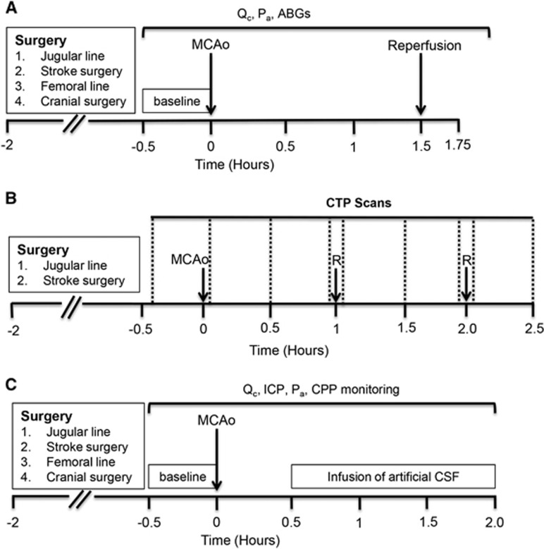
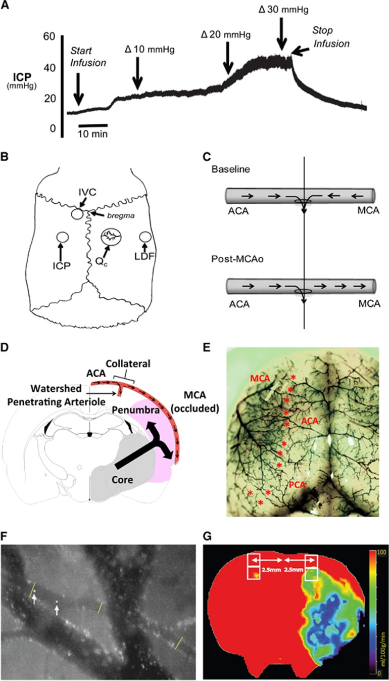
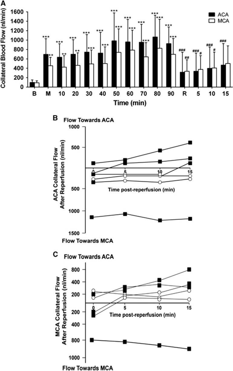
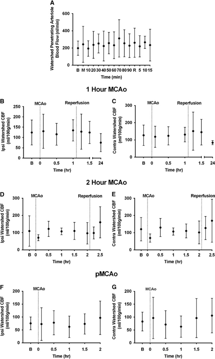
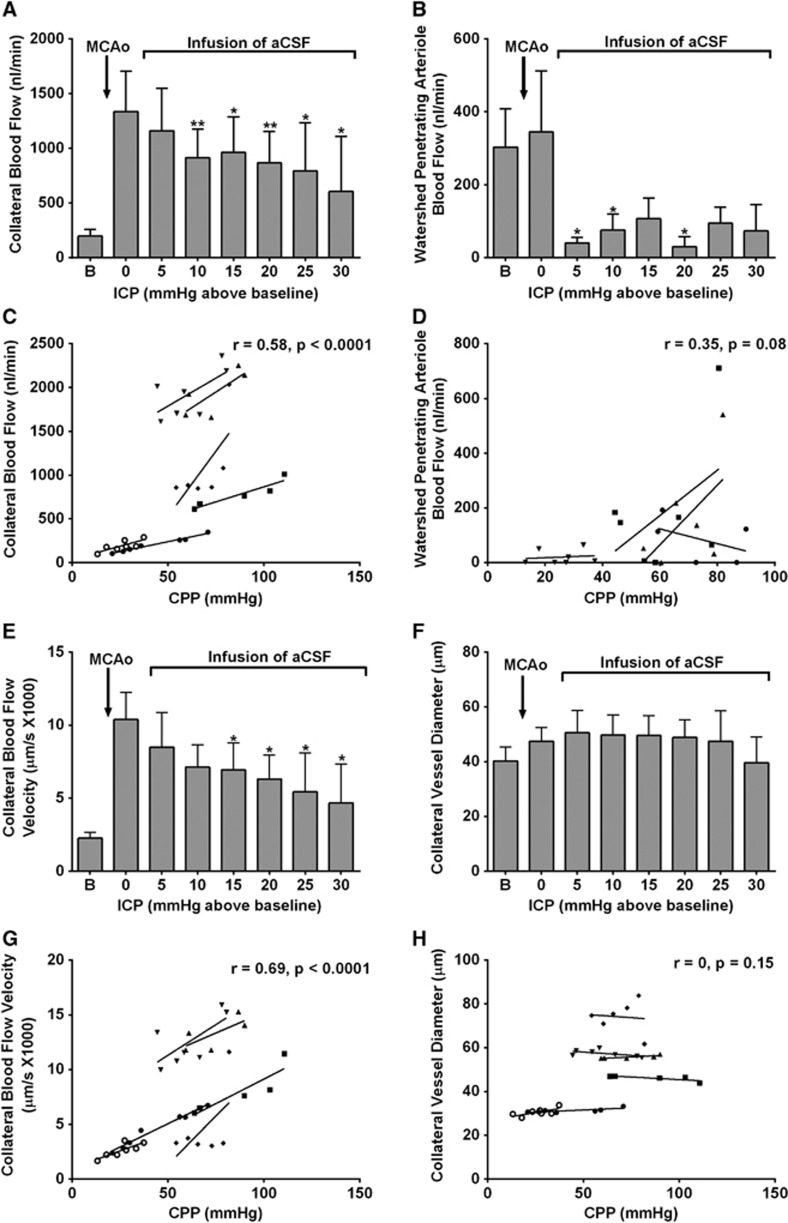
Recent human imaging studies indicate that reduced blood flow through pial collateral vessels ('collateral failure') is associated with late infarct expansion despite stable arterial occlusion. The cause for 'collateral failure' is unknown. We recently showed that intracranial pressure (ICP) rises dramatically but transiently 24 hours after even minor experimental stroke. We hypothesized that ICP elevation would reduce collateral blood flow. First, we investigated the regulation of flow through collateral vessels and the penetrating arterioles arising from them during stroke reperfusion. Wistar rats were subjected to intraluminal middle cerebral artery (MCA) occlusion (MCAo). Individual pial collateral and associated penetrating arteriole blood flow was quantified using fluorescent microspheres. Baseline bidirectional flow changed to MCA-directed flow and increased by >450% immediately after MCAo. Collateral diameter changed minimally. Second, we determined the effect of ICP elevation on collateral and watershed penetrating arteriole flow. Intracranial pressure was artificially raised in stepwise increments during MCAo. The ICP increase was strongly correlated with collateral and penetrating arteriole flow reductions. Changes in collateral flow post-stroke appear to be primarily driven by the pressure drop across the collateral vessel, not vessel diameter. The ICP elevation reduces cerebral perfusion pressure and collateral flow, and is the possible explanation for 'collateral failure' in stroke-in-progression.


Figures

Figure 1
Figure 1
Experimental timelines. (A) Study I. Collateral and ‘watershed' penetrating arteriole blood flow was measured during 90 minutes MCAo and 15 minutes reperfusion. (B) Study II. CTP imaging (vertical dotted lines) was performed to measure whole brain perfusion changes during both permanent and temporary (1 hours or 2 hours) MCAo. After baseline CTP scans, scans were taken every 30 minutes during MCAo. Further scans were taken immediately after reperfusion (R) and at 30 minutes after reperfusion. Images obtained from these scans were then used to assess changes in ‘watershed' perfusion during occlusion and reperfusion. (C) Study III. ICP was artificially elevated by fluid infusion into the lateral ventricle and effects on collateral blood flow were determined. ABG, arterial blood gas; CPP, cerebral perfusion pressure; CTP, computed tomography perfusion; ICP, intracranial pressure; MCAo, middle cerebral artery occlusion; Pa, arterial pressure; Qc, collateral blood flow.
Figure 2
Figure 2
Surgical and experimental procedures. (A) Representative intracranial pressure (ICP) trace. ICP was increased by infusion of artificial cerebrospinal fluid (aCSF) into the left lateral ventricle of a rat post-stroke. The infusion rate was increased stepwise from 4 μL/min to 40 μL/min. This resulted in an increase in ICP with no significant change in arterial blood pressure, so cerebral perfusion pressure (CPP) (not shown) mirrored ICP changes. (B) Schematic of skull surgery and monitoring: cranial window used to measure collateral flow (Qc), laser Doppler flow (LDF) probe to measure middle cerebral artery (MCA) perfusion, intraventricular catheter (IVC) for infusion of aCSF and ICP probe location. (C) Schematic of collateral blood flow direction before and after MCA occlusion (MCAo). The ‘watershed' penetrating arteriole at the confluence of the bidirectional collateral vessel blood flow (top panel) was used to demarcate the anterior (ACA) and middle (MCA) cerebral artery portions of the vessel (vertical line). This same landmark was also used after MCAo (bottom panel). Blood flow was measured in both ACA and MCA portions of the vessel at each imaging time point. (D) Schematic showing the relationship of the collateral and 'watershed' penetrating arteriole vessels in relation to infarct core and penumbra in the MCA territory at 2.3 mm behind bregma (corresponding to the center of the cranial window). Arrows within the blood vessel indicate direction of blood flow during MCAo. The large black arrow indicates direction of infarct expansion into the penumbra toward the collateral vessel. Area of infarction is based of infarct probability maps 24 hours after permanent MCAo in our model. (E) Rat cortical vasculature showing the location of pial collateral vessels (colored latex perfusion). Collaterals between the ACA and the MCA, and posterior cerebral artery and MCA are marked with asterisks. (F) Calculation of collateral vessel flow. Image shows two merged frames, taken 3.3 ms apart. A collateral vessel and larger bridging veins are seen. Arrows show the location of a microsphere at two time points—distance travelled between frames is used to calculate velocity; lines show locations of vessel diameter measurements. (G) Representative computed tomography perfusion (CTP) cerebral blood flow (CBF) map at bregma. Two regions of interest (ROIs) were fitted (white boxes) 2.5 mm lateral to midline in each hemisphere, corresponding to the location of the penetrating arterioles that are supplied by the collaterals imaged in study I. Perfusion was measured by taking the average CBF within these ROIs.
Figure 3
Figure 3
Collateral flow increases during ischemia and decreases during reperfusion. (A) Collateral blood flow: absolute blood flow in the anterior cerebral artery (ACA) segment of the collateral vessel (solid columns) and blood flow in the middle cerebral artery (MCA) segment of the collateral vessel (hollow columns) during MCA occlusion (M) and reperfusion (R); **P<0.01, ***P<0.001 for post-RM-ANOVA Dunnett's test compared with baseline (B); #P<0.05, ##P<0.01, ###P<0.001 for post-RM-ANOVA Dunnett's test compared with 90 minutes MCAo (n=6). Average blood flow is represented irrespective of direction. Flow direction varied after reperfusion, therefore averaging both + and − flows would leave the impression that there was little to no flow through these vessels, which was not the case. Panels (B and C) illustrate the importance of directionality of flow post-reperfusion in the ACA and MCA segments of the collateral vessel. (B) Individual animal analysis of direction and magnitude of blood flow through the ACA segment of collateral vessels during reperfusion (nL/min). Squares=unidirectional flow, Circles=bidirectional flow. (C) Individual animal analysis of direction and magnitude of blood flow through the MCA segment of collateral vessels during reperfusion (nL/min). Squares, unidirectional flow; circles, bidirectional flow.
Figure 4
Figure 4
Collateral flow change is strongly correlated with blood flow velocity changes and only weakly correlated with vessel diameter. (A) Collateral blood flow velocity and (B) Collateral vessel diameter, in the anterior cerebral artery (ACA, solid columns) and middle cerebral artery (MCA, hollow columns) segments of the collateral vessels during MCA occlusion (MCAo) and reperfusion. *P<0.05, **P<0.01, ***P<0.001 for post-RM-ANOVA Dunnett's test compared with baseline; #P<0.05, ##P<0.01 for post-RM-ANOVA Dunnett's test compared with 90 minutes MCAo. Repeated measures linear regression analysis of collateral blood flow versus blood flow velocity (C) and vessel diameter (D). Data points include measurements from both the ACA and MCA sides of each collateral vessel, recorded at each time point. Separate data symbols and regression lines are shown for each animal (n=6). B, baseline; M, MCAo; R, reperfusion.
Figure 5
Figure 5
‘Watershed' penetrating arteriole blood flow and ‘watershed'-region tissue perfusion remain constant throughout middle cerebral artery (MCA) occlusion (MCAo) and reperfusion. (A) Blood flow in the ‘watershed' penetrating arterioles arising from collateral vessels calculated using fluorescent microspheres. (BG) Ipsilateral and contralateral 'watershed' tissue perfusion assessed using serial computed tomography perfusion (CTP) cerebral blood flow (CBF) during MCA occlusion±reperfusion, during 1-hour MCAo (B and C), 2-hour MCAo (D and E) and permanent MCAo (pMCAo) (F and G). B, baseline; M, MCAo; R, reperfusion.
Figure 6
Figure 6
Artificial elevation of intracranial pressure (ICP) reduces both collateral and ‘watershed' penetrating arteriole flow. The reduction in collateral flow is primarily driven by a significant reduction in blood flow velocity in the collateral vessel, not changes in vessel diameter. (A) Collateral blood flow and (B) ‘Watershed' penetrating arteriole flow, at baseline and at each incremental increase of ICP. *P<0.05, **P<0.01 for post-RM-ANOVA Dunnett's test compared with post-MCAo baseline. Linear regression analysis of cerebral perfusion pressure (CPP) versus: (C) collateral blood flow, and (D) ‘Watershed' penetrating arteriole blood flow, during ICP elevation; (E) collateral blood flow velocity and (F) collateral vessel diameter, at baseline and at each incremental increase of ICP. *P<0.05, for post-RM-ANOVA Dunnett's test compared with post-MCAo baseline. Linear regression analysis of CPP versus: (G) collateral blood flow velocity and (H) collateral vessel diameter, during ICP elevation (data points are measurements recorded at each time point, n=6 animals for collateral flow, blood flow velocity, and vessel diameter; n=4 animals for penetrating arteriole flow, individual regression lines are shown). aCSF, artificial cerebrospinal fluid; MCAo, middle cerebral artery occlusion.

References

    1. Davalos A, Castillo J.Progressing strokeFisher M, Bogousslavsky J. Current Review of Cerebrovascular Disease Philadelphia: Springer; 2001pp 169–181.
    1. Ali LK, Saver JL. The ischemic stroke patient who worsens: new assessment and management approaches. Rev Neurol Dis. 2007;4:85–91. - PubMed
    1. Coutts SB, Modi J, Patel SK, Aram H, Demchuk AM, Goyal M, et al. What causes disability after transient ischemic attack and minor stroke?: Results from the CT and MRI in the Triage of TIA and minor Cerebrovascular Events to Identify High Risk Patients (CATCH) Study. Stroke. 2012;43:3018–3022. - PubMed
    1. Caplan LR. Treatment of "progressive" stroke. Stroke. 1991;22:694–695. - PubMed
    1. Roden-Jullig A, Britton M. Effectiveness of heparin treatment for progressing ischaemic stroke: before and after study. J Intern Med. 2000;248:287–291. - PubMed