. 2015 Mar 10;6(7):4992-5004.
doi: 10.18632/oncotarget.3214.

The heparan sulfate mimetic PG545 interferes with Wnt/β-catenin signaling and significantly suppresses pancreatic tumorigenesis alone and in combination with gemcitabine

Affiliations

The heparan sulfate mimetic PG545 interferes with Wnt/β-catenin signaling and significantly suppresses pancreatic tumorigenesis alone and in combination with gemcitabine

Deok-Beom Jung et al. Oncotarget. .

Erratum in

Abstract

The heparan sulfate mimetic PG545 has been shown to exert anti-angiogenic and anti-metastatic activity in vitro and in vivo cancer models. Although much of this activity has been attributed to inhibition of heparanase and heparan sulfate-binding growth factors, it was hypothesized that PG545 may additionally disrupt Wnt signaling, an important pathway underlying the malignancy of pancreatic cancer. We show that PG545, by directly interacting with Wnt3a and Wnt7a, inhibits Wnt/β-catenin signaling leading to inhibition of proliferation in pancreatic tumor cell lines. Additionally, we demonstrate for the first time that the combination of PG545 with gemcitabine has strong synergistic effects on viability, motility and apoptosis induction in several pancreatic cell lines. In an orthotopic xenograft mouse model, combination of PG545 with gemcitabine efficiently inhibited tumor growth and metastasis compared to single treatment alone. Also, PG545 treatment alone decreased the levels of β-catenin and its downstream targets, cyclin D1, MMP-7 and VEGF which is consistent with our in vitro data. Collectively, our findings suggest that PG545 exerts anti-tumor activity by disrupting Wnt/β-catenin signaling and combination with gemcitabine should be considered as a novel therapeutic strategy for pancreatic cancer treatment.

Keywords: PG545; Wnt/β-catenin; gemcitabine; heparan sulfate mimetic; pancreatic cancer.


Conflict of interest statement

Conflict of Interests

The authors disclose no potential conflicts of interest with the exception of EH and KD who are full time employees of Progen Pharmaceuticals Ltd.

Figures

Figure 1
Figure 1. Effects of PG545 on Wnt/β-catenin signaling in pancreatic cancer cells
(A) AsPC-1 cells were treated with various concentrations of PG545 for 6 h. (B) AsPC-1 cells were treated with various concentrations of PG545 for 6 h with or without 50 ng/ml of Wnt3a and subjected to western blotting. (C) AsPC-1 cells were incubated with PG545 (30 μM) and/or Wnt3a (100 ng/mL) for 24 h and the mRNA expressions of β-catenin downstream target genes were analyzed by Real-Time quantitative RT-PCR. (D) AsPC-1 cells containing β-catenin binding promoter fused with luciferase gene or control cells were incubated with PG545 for 24 h. ***P < 0.001 vs. TOPflash without PG545.
Figure 2
Figure 2. Disruption of HS-mediated Wnt/β-catenin signaling by PG545
(A) Inhibition of Wnt-heparin binding by PG545. Representative response curves for PG545 inhibition of Wnt3a and Wnt7a binding to immobilized heparin. Multiple response curves were used to determine IC50 values of 1.91 ± 0.09 nM for Wnt3a (n = 5) and 0.97 ± 0.12 nM for Wnt7a (n = 6). (B) Blocking effect of PG545 at the binding between recombinant Wnt and AsPC-1 cells was assessed by immunofluorescence. AsPC-1 cells were incubated with PG545 (20 μM) and/or recombinant His-tagged Wnt3a or Wnt7a (200 ng/mL) for 6 h as shown in MATERIALS AND METHODS. Scale bars; 5 μm. (C) Flow cytometric analysis for detection of FITC positive cells was used to observe the inhibition by PG545 of Wnt3a binding to Capan-1 and AsPC-1 cells. Cells were incubated with or without PG545 (20 μM) and/or recombinant His-tagged Wnt3a (100 ng/mL) for 2 h at 4°C. Y-axis represents the % of median FITC intensity indicating binding of Wnt ligands to cell surface. (D) Dose-dependent response for β-catenin transcriptional activation with increasing concentrations of PG545. AsPC-1 reporter and control cells were incubated with Wnt3a (100 ng/ml) and the indicated concentrations of PG545 for 24 h. ***P < 0.001 vs. TOPflash + Wnt3a without PG545.
Figure 3
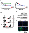
Figure 3. Effects of PG545 and gemcitabine treatment on pancreatic cancer cells
(A) AsPC-1, BxPC-3, MiaPaCa-2, and Panc-1 cells were treated with various concentrations of gemcitabine (0.05 to 0.6 μM) for 72 h or PG545 (5 to 100 μM) for 24 h. Cell viability was measured by MTT assay. (B) AsPC-1 and Panc-1 cells were treated with PBS (control), gemcitabine (50 ng/ml) or PG545 (40 μM). After staining with Annexin V-FITC and PI, the apoptotic cells were analyzed by flow cytometer. The numbers in each plot indicate the percentage of apoptotic cells, i.e. positive staining for both Annexin V and PI. (C) The cells treated with PG545 (20 μM) for 6 h were subjected to Western blotting. Experimental conditions of (D) using Panc-1 cells were same in Figure 2B. Scale bars; 5 μm.
Figure 4
Figure 4. Synergistic effects of PG545 and gemcitabine co-treatment on pancreatic cancer cells
(A) AsPC-1, BxPC-3, MiaPaCa-2, and Panc-1 cells were placed on 96-well plates and treated with gemcitabine (0.05~0.6 μM) alone or in combination with 5 or 10 μM of PG545. Cell viability was measured by MTT assay. Data represent means ± S.D. from three independent experiments. (B) Combination index (CI) values with fraction affected (Fa) between gemcitabine and PG545 in AsPC-1, BxPC-3, MiaPaCa-2 and Panc-1 cells was calculated using Calcusyn software. (C) and (D) AsPC-1 cells were treated with indicated concentrations of PG545 and gemcitabine for 48h. (C) The other procedures of apoptosis assay are same in Fig. 3B. (D) Cell lysates were subjected to Western blotting.
Figure 5
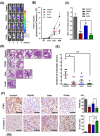
Figure 5. Inhibition of tumor growth and metastasis by PG545 in AsPC-1 orthotopic xenograft mouse model
Treatment groups: Control group data, grey or black symbols and bars; PG545 group data, red; Gem group data, blue; Combination group (at half the PG545 and Gem doses) data, green. See Materials and Methods for experimental details. (A) Bioluminescence IVIS images of orthotopically implanted pancreatic tumors in live and anesthetized mice (n=7-8). (B) Tumor volume during the experiment. (C) Final tumor weight at termination of experiment. Tumor data expressed relative to average tumor weight of control group. **P < 0.01, ***P < 0.001 vs. control; #P < 0.025 vs. PG545 alone. (D) Lung tissue sections were stained with Hematoxylin and Eosin. (E) Number of lung metastases. *P < 0.05, **P < 0.01. (F) Representative examples of immunohistochemical staining for PCNA and Cleaved Caspase-3 in tumor sections with histograms showing quantitation of staining.
Figure 6
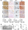
Figure 6. PG545 inhibits β-catenin signaling in AsPC-1 orthotopic xenograft mouse model
(A) Immunohistochemical analysis of β-catenin and β-catenin-regulated proteins, Cyclin D1, MMP-7 and VEGF in pancreatic tumor tissues from mice. Quantitation of staining was performed using 10 fields per analyte. **P < 0.01, ***P < 0.001. (B) Frozen tumor tissues were homogenized on ice and the extracts were subjected to Western blotting. (C) β-catenin and (D) Cyclin D1 levels were quantified by Image J software and plotted relative to the control group in Fig. 6B. **P < 0.01, ***P < 0.001 vs. control.

References

    1. Jemal A, Siegel R, Ward E, Murray T, Xu J, Thun MJ. Cancer statistics, 2007. CA Cancer J Clin. 2007;57(1):43–66. - PubMed
    1. Ghaneh P, Costello E, Neoptolemos JP. Biology and management of pancreatic cancer. Gut. 2007;56(8):1134–1152. - PMC - PubMed
    1. Di Marco M, Di Cicilia R, Macchini M, Nobili E, Vecchiarelli S, Brandi G, Biasco G. Metastatic pancreatic cancer: is gemcitabine still the best standard treatment? (Review) Oncol Rep. 2010;23(5):1183–1192. - PubMed
    1. Wang L, Heidt DG, Lee CJ, Yang H, Logsdon CD, Zhang L, Fearon ER, Ljungman M, Simeone DM. Oncogenic function of ATDC in pancreatic cancer through Wnt pathway activation and beta-catenin stabilization. Cancer Cell. 2009;15(3):207–219. - PMC - PubMed
    1. Gui S, Yuan G, Wang L, Zhou L, Xue Y, Yu Y, Zhang J, Zhang M, Yang Y, Wang DW. Wnt3a regulates proliferation, apoptosis and function of pancreatic NIT-1 beta cells via activation of IRS2/PI3K signaling. J Cell Biochem. 2013;114(7):1488–1497. - PubMed

Publication types

MeSH terms