. 2015 Apr 3;290(14):8904-12.
doi: 10.1074/jbc.M114.612796. Epub 2015 Feb 10.

Ubiquitinated sirtuin 1 (SIRT1) function is modulated during DNA damage-induced cell death and survival

Affiliations

Ubiquitinated sirtuin 1 (SIRT1) function is modulated during DNA damage-induced cell death and survival

Lirong Peng et al. J Biol Chem. .

Abstract

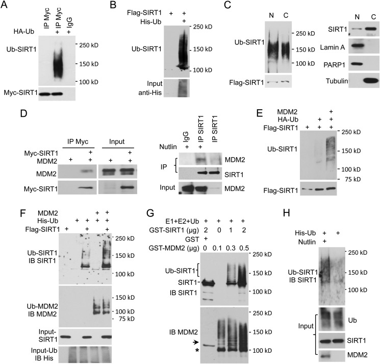
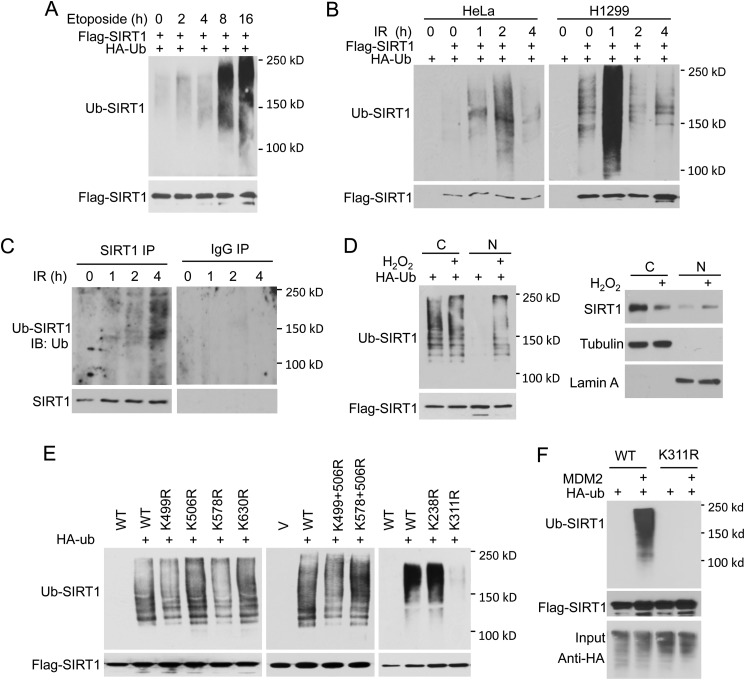
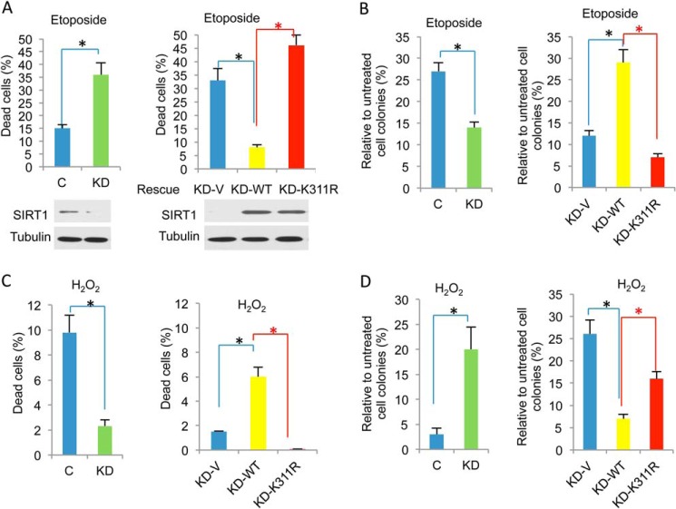
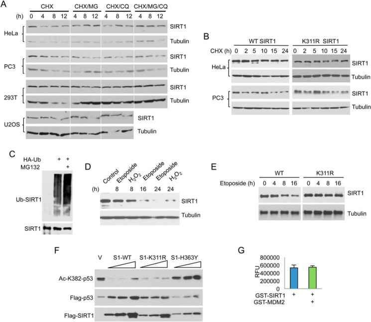
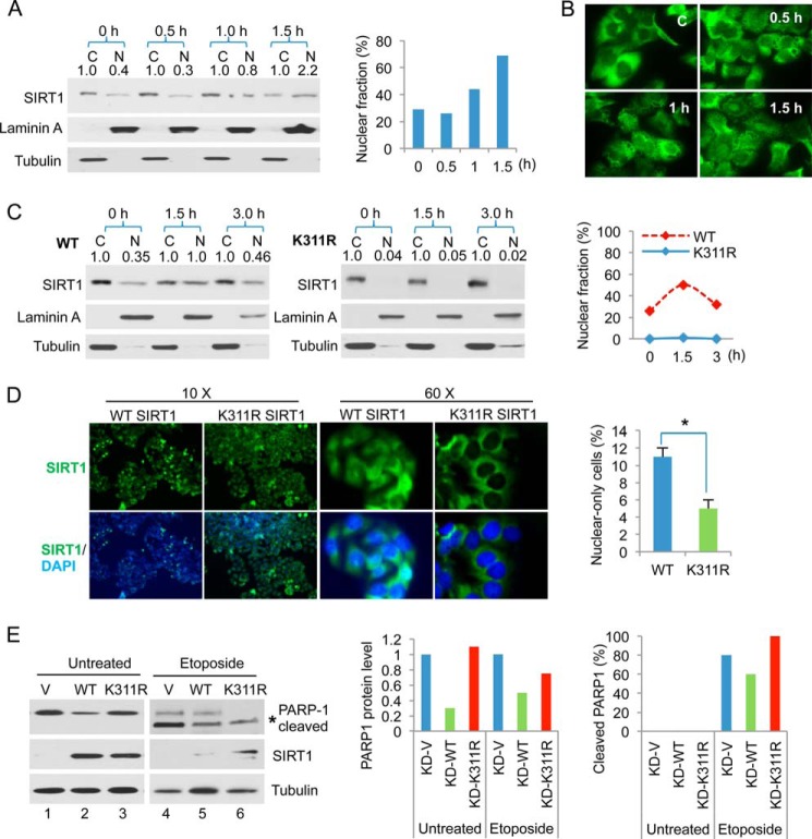
Downstream signaling of physiological and pathological cell responses depends on post-translational modification such as ubiquitination. The mechanisms regulating downstream DNA damage response (DDR) signaling are not completely elucidated. Sirtuin 1 (SIRT1), the founding member of Class III histone deacetylases, regulates multiple steps in DDR and is closely associated with many physiological and pathological processes. However, the role of post-translational modification or ubiquitination of SIRT1 during DDR is unclear. We show that SIRT1 is dynamically and distinctly ubiquitinated in response to DNA damage. SIRT1 was ubiquitinated by the MDM2 E3 ligase in vitro and in vivo. SIRT1 ubiquitination under normal conditions had no effect on its enzymatic activity or rate of degradation; hypo-ubiquitination, however, reduced SIRT1 nuclear localization. Ubiquitination of SIRT1 affected its function in cell death and survival in response to DNA damage. Our results suggest that ubiquitination is required for SIRT1 function during DDR.

Keywords: Deubiquitylation (Deubiquitination); Histone Deacetylase (HDAC); Post-translational Modification (PTM); Sirtuin; Ubiquitylation (Ubiquitination).


Figures

FIGURE 1.
FIGURE 1.
SIRT1 is ubiquitinated. A, 293T cells were transfected with Myc-SIRT1 and either HA-Ub or HA-tag control vector. After 36 h, cells were treated with 50 μm MG132 for 4 h. SIRT1 was immunoprecipitated (IP) using anti-Myc antibody and IgG isotype control and then analyzed for ubiquitination by Western blot using anti-HA antibody. B, cells were transfected with FLAG-SIRT1 and either His-Ub or control vector. Ubiquitinated protein was pulled down by NTA+ beads and resolved by SDS-PAGE. SIRT1 in the pulldown fraction was detected by anti-SIRT1 antibody. C, 293T cells were transfected with FLAG-SIRT1 and HA-Ub, and then cytoplasmic (C) and nuclear (N) extracts were prepared. Expression levels of SIRT1 in cellular fractions were examined by Western blot (right). FLAG-SIRT1 was immunoprecipitated using anti-FLAG antibody. Equal amounts of SIRT1 from both fractions were loaded for ubiquitination analysis (left). D, interaction between MDM2 and SIRT1. 293T cells were co-transfected with Myc-SIRT1 and MDM2. SIRT1 was immunoprecipitated using anti-Myc antibody. Co-immunoprecipitation of MDM2 was detected using anti-MDM2 antibody (left). A549 cells were treated with 7 μm Nutlin overnight or left untreated. Endogenous SIRT1 was pulled down by anti-SIRT1 antibody, and MDM2 co-immunoprecipitation was detected using anti-MDM2 antibody (right). E, SIRT1 is ubiquitinated by MDM2 in vivo. FLAG-SIRT1 was co-expressed with HA-Ub and either MDM2 or vector control. SIRT1 was immunoprecipitated by anti-FLAG antibody and analyzed for ubiquitination using anti-HA antibody. F, FLAG-SIRT1 was co-expressed with His-Ub and MDM2 or control vector. SIRT1 ubiquitination was analyzed as described in B. IB, immunoblot. G, SIRT1 was ubiquitinated by MDM2 in vitro. GST-SIRT1 was incubated with active E1, E2, and ubiquitin in the presence of GST-MDM2 or GST alone. Reaction products were analyzed by Western blot using anti-SIRT1 antibody. The membrane was stripped and re-probed with anti-MDM2 antibody. The arrow indicates residual SIRT1 signal after stripping. The asterisk (*) indicates the band corresponding to non-ubiquitinated GST-MDM2. H, A549 cells were transfected with His-Ub. At 36 h after transfection, cells were treated with 7 μm Nutlin overnight and 50 μm MG132 for 4 h before harvesting. His-Ub-conjugated proteins were pulled down, and ubiquitination of endogenous SIRT1 was examined as described in F.
FIGURE 2.
FIGURE 2.
SIRT1 ubiquitination is regulated by DNA damage. A–D, DNA damage regulates SIRT1 ubiquitination. FLAG-SIRT1 was co-transfected with HA-Ub. At 36 h post-transfection, cells were treated with 50 μm etoposide, 10 gray IR, or left untreated. FLAG-SIRT1 was immunoprecipitated using anti-FLAG antibody and analyzed for ubiquitination using anti-HA antibody. C, ubiquitination of endogenous SIRT1. Endogenous SIRT1 was immunoprecipitated from HeLa cells using anti-SIRT1 antibody, and ubiquitination was assessed using anti-ubiquitin antibody. D, HeLa cells were transfected with FLAG-SIRT1 and HA-Ub. At 36 h after transfection, cells were treated with 400 μm H2O2 or left untreated. Cytoplasmic (C) and nuclear (N) fractions were extracted and examined for SIRT1 expression (right). Cytoplasmic and nuclear fractions were normalized to contain the same amount of SIRT1, used for immunoprecipitation, and then examined for ubiquitination (left). E and F, WT and lysine-to-arginine mutant of SIRT1 were transfected with or without HA-Ub and along with MDM2 or not. SIRT1 ubiquitination was analyzed as described in Fig. 1A. V, vector control.
FIGURE 3.
FIGURE 3.
K311R is defective in regulating cell death and survival during DDR. A, control and SIRT1 KD HeLa cells were treated with 5 μm etoposide for 24 h (left). SIRT1 KD HeLa cells were transfected with WT SIRT1, K311R SIRT1, or vector control (C) and subsequently treated with etoposide (right). Apoptosis and necrosis were examined by flow cytometric analysis using annexin V-PI staining. The percentage of dead cells was determined by measuring the percentage of annexin-positive cells. Levels of SIRT1 and tubulin were assessed by Western blot. B, control, KD, and rescued cells were treated with 5 μm etoposide for 2 h or left untreated. Two weeks later cell colonies were counted and normalized to the untreated control. C, control and SIRT1 KD HeLa cells were treated with 100 μm H2O2 for 2 h (left). SIRT1 KD HeLa cells were transfected with WT SIRT1, K311R SIRT1, or vector control and subsequently treated with H2O2 (right). Cell death was measured by annexin V-PI staining. D, control, KD, and rescued cells were treated with 100 μm H2O2 for 30 min or left untreated. Two weeks later, cell colonies were counted and normalized to the untreated control. All data are presented as the means ± S.E. of at least three independent experiments. A two-tailed unpaired Student's t test was applied to calculate the p value using the GraphPad Prism software. *, p < 0.05 was considered as statistically significant.
FIGURE 4.
FIGURE 4.
SIRT1 stability is regulated by DNA damage. A and B, various cell lines were treated with 50 μg/ml cycloheximide (CHX) alone or together with 50 μm MG132 (MG), 20 μm chloroquine (CQ), or both for the indicated times or left untreated. SIRT1 and tubulin protein levels were examined by Western blot. C, ubiquitination of endogenous SIRT1 in the presence or absence of MG132. D, cells were treated with etoposide and H2O2 for the indicated times, and SIRT1 protein level was assessed by Western blot. E, WT and K311R stably transfected HeLa cells were treated with 50 μm etoposide for the indicated times. Protein levels of WT and K311R SIRT1 were examined by Western blot using anti-SIRT1 antibody. F, SIRT1 deacetylase activity in vivo. HeLa cells were co-transfected with FLAG-p53 and a vector control (V) or an increasing amount of WT, K311R, and H353Y SIRT1. Acetylation of p53 at K382 was examined by Western blot using anti-Ac-K382 p53 antibody. Then, the membrane was stripped and reprobed with anti-FLAG antibodies. G, SIRT1 deacetylase activity in vitro. GST-SIRT1 was first ubiquitinated by GST-MDM2 in vitro as described in Fig. 1G. An aliquot of the reaction mixture (containing 0.5 μg SIRT1) was incubated with 100 μm acetylated p53 peptides and 3 mm NAD+ for 1 h at 32 °C. Fluorescence was measured using a fluorometric plate reader, and is presented as relative fluorescence units (RFU).
FIGURE 5.
FIGURE 5.
SIRT1 ubiquitination affects subcellular localization. A, DU145 cells were treated with 10 gray IR for the indicated times, and then nuclear (N) and cytoplasmic (C) extracts were prepared for Western blot analysis using antibodies specific for SIRT1, lamin A (nuclear marker), and tubulin (cytoplasmic marker). SIRT1 protein levels were determined by densitometry and are indicated for each fraction. The percentage of SIRT1 in the nucleus relative to total SIRT1 was calculated using the values determined by densitometry. B, DU145 cells were treated as above, and subcellular localization of endogenous SIRT1 was examined by immunofluorescence. C, HeLa cells expressing WT or K311R SIRT1 were treated as in A. The percentage of SIRT1 present in the nucleus was quantified relative to total SIRT1. D, immunofluorescence analysis of SIRT1 localization in WT and K311R-rescued HeLa cells. Nuclei were counterstained with DAPI. 10× and 60× images are shown. The percentage of cells containing only nuclear-localized SIRT1 was calculated and compared between WT and K311R cells. *, p < 0.002. E, SIRT1-KD cells were rescued with WT, K311R, or vector (V) control and treated with 20 μm etoposide for 24 h or left untreated. Because WT SIRT1 rescue substantially reduced PARP-1, 3-fold more WT cell lysate was loaded onto the fifth lane to clearly show PARP-1 cleavage. Total and cleaved PARP-1, SIRT1, and tubulin were examined by Western blot (left). PARP-1 protein levels in WT and K311R SIRT1 cells were normalized with respect to the vector control (middle). The percentage of PARP-1 cleavage was quantified relative to total PARP-1 (right).

References

    1. Peng L., Seto E. (2011) Deacetylation of nonhistone proteins by HDACs and the implications in cancer. Handb. Exp. Pharmacol. 206, 39–56 - PubMed
    1. Finkel T., Deng C. X., Mostoslavsky R. (2009) Recent progress in the biology and physiology of sirtuins. Nature 460, 587–591 - PMC - PubMed
    1. Satoh A., Stein L., Imai S. (2011) The role of mammalian sirtuins in the regulation of metabolism, aging, and longevity. Handb. Exp. Pharmacol. 206, 125–162 - PMC - PubMed
    1. Haigis M. C., Guarente L. P. (2006) Mammalian sirtuins–emerging roles in physiology, aging, and calorie restriction. Genes Dev. 20, 2913–2921 - PubMed
    1. Haigis M. C., Sinclair D. A. (2010) Mammalian sirtuins: biological insights and disease relevance. Annu. Rev. Pathol. 5, 253–295 - PMC - PubMed

Publication types