. 2015 Feb;9(1):3-10.
doi: 10.4162/nrp.2015.9.1.3. Epub 2014 Aug 30.

Intestinal anti-inflammatory activity of Sasa quelpaertensis leaf extract by suppressing lipopolysaccharide-stimulated inflammatory mediators in intestinal epithelial Caco-2 cells co-cultured with RAW 264.7 macrophage cells

Affiliations

Intestinal anti-inflammatory activity of Sasa quelpaertensis leaf extract by suppressing lipopolysaccharide-stimulated inflammatory mediators in intestinal epithelial Caco-2 cells co-cultured with RAW 264.7 macrophage cells

Kyung-Mi Kim et al. Nutr Res Pract. 2015 Feb.

Abstract

Background/objectives: Inflammatory bowel disease (IBD), including Crohn's disease and ulcerative colitis, involves chronic inflammation of the gastrointestinal tract. Previously, Sasa quelpaertensis leaves have been shown to mediate anti-inflammation and anti-cancer effects, although it remains unclear whether Sasa leaves are able to attenuate inflammation-related intestinal diseases. Therefore, the aim of this study was to investigate the anti-inflammatory effects of Sasa quelpaertensis leaf extract (SQE) using an in vitro co-culture model of the intestinal epithelial environment.

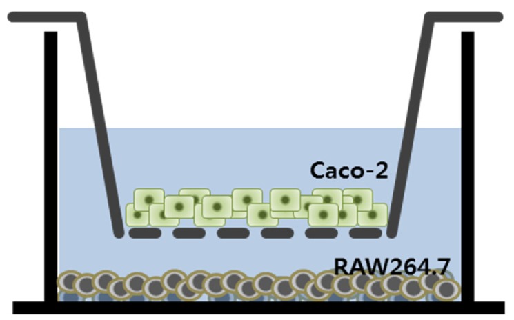
Materials/methods: An in vitro co-culture system was established that consisted of intestinal epithelial Caco-2 cells and RAW 264.7 macrophages. Treatment with lipopolysaccharide (LPS) was used to induce inflammation.

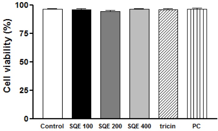
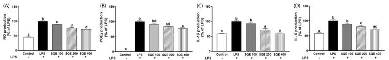
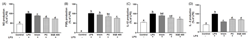
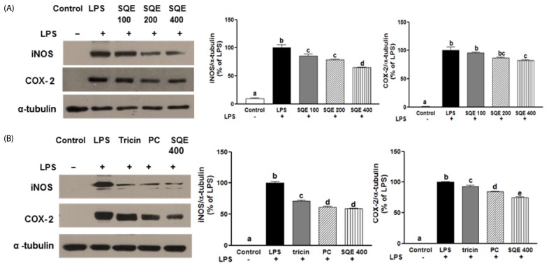
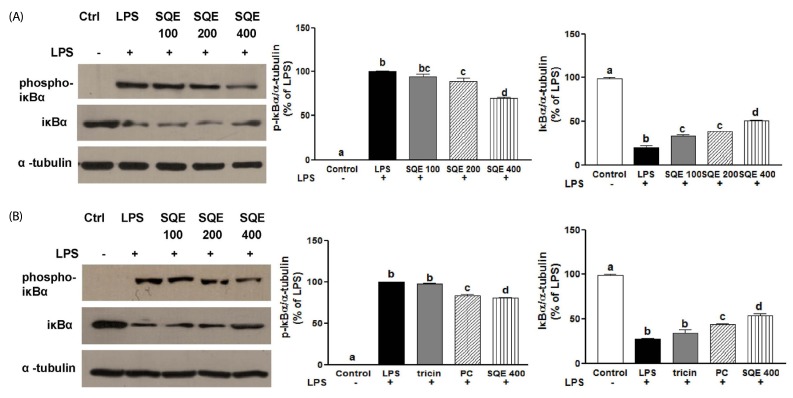
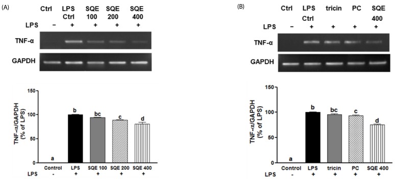
Results: Treatment with SQE significantly suppressed the secretion of LPS-induced nitric oxide (NO), prostaglandin E2 (PGE2), IL-6, and IL-1β in co-cultured RAW 264.7 macrophages. In addition, expressions of inducible nitric oxide synthase (iNOS), cyclooxygenase (COX)-2, and tumor necrosis factor (TNF)-α were down-regulated in response to inhibition of IκBα phosphorylation by SQE. Compared with two bioactive compounds that have previously been identified in SQE, tricin and P-coumaric acid, SQE exhibited the most effective anti-inflammatory properties.

Conclusions: SQE exhibited intestinal anti-inflammatory activity by inhibiting various inflammatory mediators mediated through nuclear transcription factor kappa-B (NF-kB) activation. Thus, SQE has the potential to ameliorate inflammation-related diseases, including IBD, by limiting excessive production of pro-inflammatory mediators.

Keywords: Sasa quelpaertensis; anti-inflammation; co-culture model; pro-inflammatory mediators.


Figures

Fig. 1
Fig. 1
Schematic representation of the in vitro co-culture system that was established. Caco-2 cells were grown in 6-well cell culture inserts with a semipermeable support membrane. Murine RAW 264.7 macrophages were cultured in 6-well culture plates. Culture inserts containing Caco-2 cells were placed in the 6-well plates to establish the co-culture system.
Fig. 2
Fig. 2
SQE did not affect the viability of RAW 264.7 macrophages in the co-culture system. RAW 264. 7 cells were seeded in 12 well-plates and treated with various doses of SQE (100, 200, and 400 µg/ml), as well as individually with two bioactive compounds, tricin (1 µM) and P-coumaric acid (PC, 2.4 µM), at concentration equivalent to that contained in 400 µg/ml SQE. After 24 h, each set of cells was stained with trypan blue. Both stained and unstained cells were counted, and the proportion of unstained cells was used to calculate cell viability. One-way ANOVA was applied using Tukey's post-hoc test (α = 0.05). SQE, Sasa quelpaertensis extract; PC, P-coumaric acid.
Fig. 3
Fig. 3
SQE reduces the expression of inflammatory mediators in the co-culture system of Caco-2 and RAW 264.7 cells. Caco-2 cells were incubated with RAW 264.7 macrophages in the presence of various concentrations of SQE (100, 200, and 400 µg/ml) that were applied to the apical side of this co-culture system. After 3 h, 1 µg/ml LPS was added to the basolateral side and the system was incubated overnight. Levels of NO (A), PGE2 (B), IL-1β (C), and IL-6 (D) were subsequently measured from collected culture medium samples using commercially available ELISA kits. Data were analyzed using one-way ANOVA for multiple comparisons followed by Tukey's post-hoc test (P < 0.05). The different letters are used to indicate significant differences. SQE, Sasa quelpaertensis extract; PC, P-coumaric acid, LPS, lipopolysaccharide.
Fig. 4
Fig. 4
Tricin and P-coumaric acid suppress the expression of inflammatory mediators in a co-culture system of Caco-2 and RAW 264.7 cells. Tricin (1 µM) and P-coumaric acid (PC, 2.4 µM) were individually added into the apical compartment of a Caco-2/RAW 264.7 co-culture model. After 3 h, 1µg/ml LPS was added to the basolateral compartment and the cells were incubated overnight. Production of NO (A), PGE2 (B), IL-1β (C), and IL-6 (D) were subsequently analyzed from collected media samples using commercially available ELISA kits. Data were analyzed by one-way ANOVA for multiple comparisons followed by Tukey's post-hoc test (P < 0.05). The different letters are used to indicate significant differences. SQE, Sasa quelpaertensis extract; PC, P-coumaric acid, LPS, lipopolysaccharide.
Fig. 5
Fig. 5
SQE, tricin, and P-coumaric acid down-regulate the expression of iNOS and COX-2 in a co-culture system of Caco-2 and RAW 264.7 cells. SQE, tricin (1 µM), and P-coumaric acid (PC, 2.4 µM) were individually added into the apical compartment of the Caco-2/RAW 264.7 co-culture model. After 3 h, 1 µg/ml LPS was added to the basolateral compartment and cells were incubated overnight. Expression of iNOS and COX-2 were then analyzed by western blot. (A) Following the addition of various doses of SQE (100, 200, 400 µg/ml), levels of iNOS and COX-2 were analyzed. The representative blots were shown (left panel) and quantified iNOS (middle panel) and COX-2 (right panel) were shown after normalization to α-tubulin. (B) A equivalent dose of tricin or PC based on 400 µg/ml SQE was added, and levels of iNOS and COX-2 were analyzed. The representative blot has shown (left panel). The quantified iNOS (middle panel) and COX-2 (right panel) were shown after normalization to α-tubulin. One-way ANOVA was applied using Tukey's post-hoc test (α = 0.05). The different letters indicate significant differences. SQE, Sasa quelpaertensis extract; PC, P-coumaric acid, LPS, lipopolysaccharide.
Fig. 6
Fig. 6
SQE, P-coumaric acid, and tricin decrease IκBα phosphorylation. SQE, tricin (1 µM), and P-coumaric acid (PC, 2.4 µM) acid were individually added into the apical compartment of the Caco-2/RAW 264.7 co-culture model. After 3 h, 1 µg/ml LPS was added to the basolateral compartment and the cells were incubated overnight. Phosphorylation of IκBα was detected by western blot. (A) Following the addition of various doses of SQE (100, 200, 400 µg/ml), levels of phospho-IκBα and IκBα were analyzed. The representative blots were shown (left panel) and quantified phospho-IκBα (middle panel) and IκBα (right panel) were presented with normalization to α-tubulin. (B) A equivalent dose of tricin or P-coumaric acid based on 400 µg/ml SQE was added, and the levels of phospho-IκBα and IκBα were analyzed. The representative blots were shown (left panel) and quantified phospho-IκBα (middle panel) and IκBα (right panel) were presented after normalization to α-tubulin. One-way ANOVA was applied using Tukey's post-hoc test (α = 0.05). The different letters indicate significant differences. SQE, Sasa quelpaertensis extract; PC, P-coumaric acid, LPS, lipopolysaccharide.
Fig. 7
Fig. 7
SQE, P-coumaric acid, and tricin down-regulate mRNA levels of TNF-α. SQE, tricin (1 µM), and P-coumaric acid (PC, 2.4 µM) were individually added into the apical compartment of the Caco-2/RAW 264.7 co-culture model. After 3 h, 1 µg/ml LPS was added to the basolateral compartment and cells were incubated overnight. TNF-α mRNA expression was analyzed using PCR. (A) Following the addition of various doses of SQE (100, 200, 400 µg/ml), levels of TNF-α mRNA were detected. The representative blots were shown (upper panel) and quantified TNF-α expression with normalization to GAPDH was presented (lower panel). (B) An equivalent dose of tricin or P-coumaric acid based on 400 µg/ml SQE was added and mRNA levels of TNF-α were detected. The representative blots has shown (upper panel) and quantified TNF-α expression with normalization to GAPDH was presented (lower panel). One-way ANOVA was applied using Tukey's post-hoc test (α = 0.05). The different letters indicate significant differences. SQE, Sasa quelpaertensis extract; PC, P-coumaric acid, LPS, lipopolysaccharide, GAPDH, glyceraldehydes-3-phosphate dehydrogenase.

References

    1. Arai F, Takahashi T, Furukawa K, Matsushima K, Asakura H. Mucosal expression of interleukin-6 and interleukin-8 messenger RNA in ulcerative colitis and in Crohn's disease. Dig Dis Sci. 1998;43:2071–2079. - PubMed
    1. Chung HL, Yue GG, To KF, Su YL, Huang Y, Ko WH. Effect of Scutellariae Radix extract on experimental dextran-sulfate sodium-induced colitis in rats. World J Gastroenterol. 2007;13:5605–5611. - PMC - PubMed
    1. Loftus EV, Jr, Sandborn WJ. Epidemiology of inflammatory bowel disease. Gastroenterol Clin North Am. 2002;31:1–20. - PubMed
    1. Bouma G, Strober W. The immunological and genetic basis of inflammatory bowel disease. Nat Rev Immunol. 2003;3:521–533. - PubMed
    1. Menditto A, Menotti A, Morisi G, Patriarca M, Spagnolo A. Serum ascorbic acid levels in men aged 55-75 years: association to selected social factors and biochemical parameters. Arch Gerontol Geriatr. 1992;15(Suppl 1):257–265. - PubMed