. 2014 Oct 16:2:17.
doi: 10.1186/2050-5736-2-17. eCollection 2014.

First noninvasive thermal ablation of a brain tumor with MR-guided focused ultrasound

Affiliations

First noninvasive thermal ablation of a brain tumor with MR-guided focused ultrasound

Daniel Coluccia et al. J Ther Ultrasound. .

Abstract

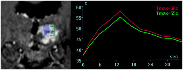
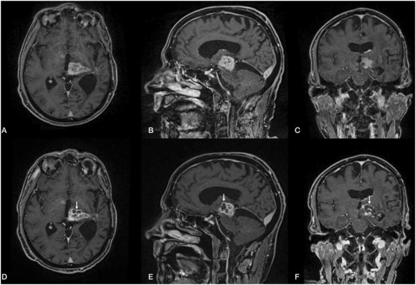
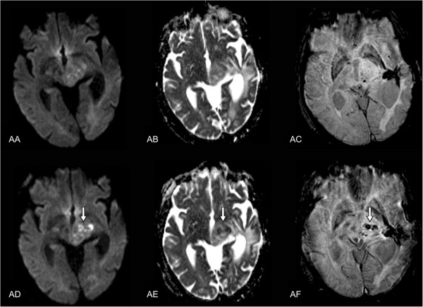
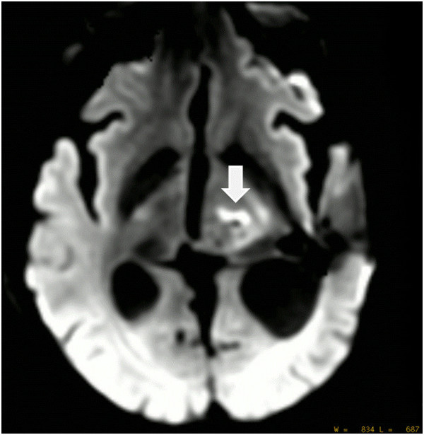
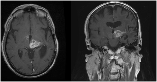
Magnetic resonance-guided focused ultrasound surgery (MRgFUS) allows for precise thermal ablation of target tissues. While this emerging modality is increasingly used for the treatment of various types of extracranial soft tissue tumors, it has only recently been acknowledged as a modality for noninvasive neurosurgery. MRgFUS has been particularly successful for functional neurosurgery, whereas its clinical application for tumor neurosurgery has been delayed for various technical and procedural reasons. Here, we report the case of a 63-year-old patient presenting with a centrally located recurrent glioblastoma who was included in our ongoing clinical phase I study aimed at evaluating the feasibility and safety of transcranial MRgFUS for brain tumor ablation. Applying 25 high-power sonications under MR imaging guidance, partial tumor ablation could be achieved without provoking neurological deficits or other adverse effects in the patient. This proves, for the first time, the feasibility of using transcranial MR-guided focused ultrasound to safely ablate substantial volumes of brain tumor tissue.

Keywords: Brain tumor; Focused ultrasound; HIFU; MRgFUS; Thermal ablation; Transcranial.


Figures

Figure 1
Figure 1
Coronal MR sequences of the tumor as depicted on the operator workstation. Console (left image). Blue marked areas correspond to completed sonication volumes; the area within the green frame illustrates the consecutively planed treatment target. Thermometric mapping (right image) shows a rapid drop of temperature within the tissue target after sonication.
Figure 2
Figure 2
Pre- (A, B, C) and post-interventional (D, E, F) MR findings. Axial, coronal, and sagittal contrast-enhanced T1-weighted, fat-saturated 3D VIBE sequence (TR = 6.2 ms; TE = 2.38 ms; flip angle = 12°; acquisition matrix = 320 × 320 pixels, section thickness = 0.9 mm) depicts a contrast-enhanced tumor with a progressive necrotic center in the post-interventional follow-up after 5 days.
Figure 3
Figure 3
Pre- (AA-AC) and post-interventional (AD-AF) MR findings. Axial diffusion weighted single-shot echoplanar imaging (A, D) (TR = 4,900 ms; TE = 130 ms; flip angle = 90°; acquisition matrix = 192 × 192 pixels, section thickness = 5 mm; spacing between slices: 6.5 mm; diffusion gradient approximately 0 and 1,000 cm2/s), corresponding ADC map (B, E), and axial flow-compensated 3D gradient-echo image (C, F) (TR = 49 ms; TE = 40 ms; flip angle = 15°; acquisition matrix = 224 × 256 pixels, section thickness = 2.0 mm) illustrate a discrete intratumoral diffusion restriction in contrast to the notable intratumoral susceptibility in the post-interventional follow-up after 5 days.
Figure 4
Figure 4
DWI image 30 min after intervention revealed significant damage to the sonicated tumor tissue. A total of 25 sonications were applied with up to 19,550 J, 17 sonications reached ablative temperatures >55°C with a maximum of 65°C.
Figure 5
Figure 5
MRI findings on day 21 after sonication of the tumor. Axial (TR = 766 ms; TE = 20 ms; acquisition matrix = 512 × 512 pixels, section thickness = 5.0 mm) and coronal (TR = 500 ms; TE = 9 ms; acquisition matrix = 512 × 512 pixels, section thickness = 5.0 mm) contrast-enhanced T1-weighted sequences image demonstrating stable findings after sonication of tumor tissue.

References

    1. Jagannathan J, Sanghvi NT, Crum LA, Yen CP, Medel R, Dumont AS, Sheehan JP, Steiner L, Jolesz F, Kassell NF. High-intensity focused ultrasound surgery of the brain: part 1–A historical perspective with modern applications. Neurosurgery. 2009;64(2):201–10. doi: 10.1227/01.NEU.0000336766.18197.8E. discussion 10–1. - DOI - PMC - PubMed
    1. Martin E, Jeanmonod D, Morel A, Zadicario E, Werner B. High-intensity focused ultrasound for noninvasive functional neurosurgery. Ann Neurol. 2009;66(6):858–61. doi: 10.1002/ana.21801. - DOI - PubMed
    1. Elias WJ, Huss D, Voss T, Loomba J, Khaled M, Zadicario E, Frysinger RC, Sperling SA, Wylie S, Monteith SJ, Druzgal J, Shah BB, Harrison M, Wintermark M. A pilot study of focused ultrasound thalamotomy for essential tremor. N Engl J Med. 2013;369(7):640–48. doi: 10.1056/NEJMoa1300962. - DOI - PubMed
    1. Lipsman N, Schwartz ML, Huang Y, Lee L, Sankar T, Chapman M, Hynynen K, Lozano AM. MR-guided focused ultrasound thalamotomy for essential tremor: a proof-of-concept study. Lancet Neurol. 2013;12(5):462–68. doi: 10.1016/S1474-4422(13)70048-6. - DOI - PubMed
    1. Jeanmonod D, Magara A, Kowalski M, Buler R, Payam P. Study of incisionless transcranial magnetic resonance-guided focused ultrasound treatment of Parkinson’s disesase: Safety, accuracy and initial outcomes 3rd International Symposium on Focused Ultrasound. Washington, DC;