

Generation of a felinized swine endothelial cell line by expression of feline decay-accelerating factor
- PMID: 25671605
- PMCID: PMC4324824
- DOI: 10.1371/journal.pone.0117682
Generation of a felinized swine endothelial cell line by expression of feline decay-accelerating factor
Abstract
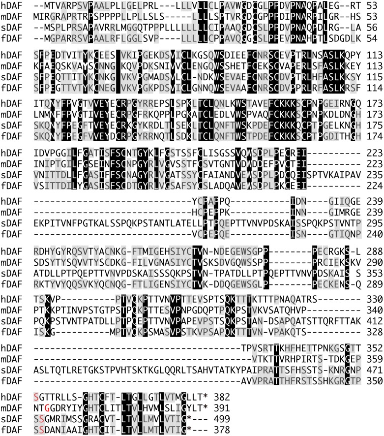
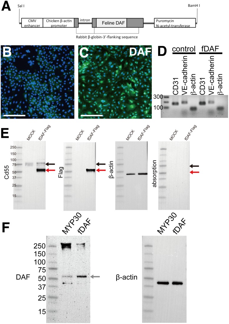
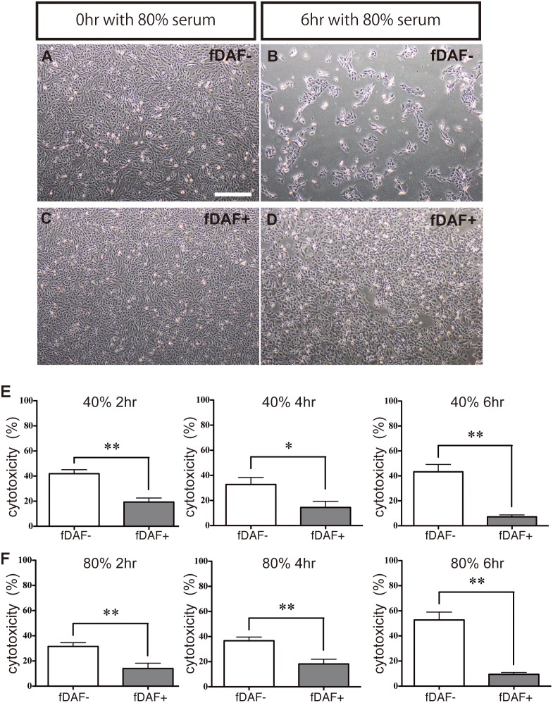
Embryonic stem cell research has facilitated the generation of many cell types for the production of tissues and organs for both humans and companion animals. Because ≥30% of pet cats suffer from chronic kidney disease (CKD), xenotransplantation between pigs and cats has been studied. For a successful pig to cat xenotransplant, the immune reaction must be overcome, especially hyperacute rejection. In this study, we isolated the gene for feline decay-accelerating factor (fDAF), an inhibitor of complement proteins, and transfected a swine endothelial cell line with fDAF to "felinize" the pig cells. These fDAF-expressing cells were resistant to feline serum containing anti-pig antibodies, suggesting that felinized pig cells were resistant to hyperacute rejection. Our results suggest that a "felinized" pig kidney can be generated for the treatment of CKD in cats in the future.
Conflict of interest statement
Figures
Similar articles
-
Expression of human decay accelerating factor may protect pig lung from hyperacute rejection by human blood.J Heart Lung Transplant. 1997 Feb;16(2):231-9. J Heart Lung Transplant. 1997. PMID: 9059935
-
Successful cross-breeding of cloned pigs expressing endo-beta-galactosidase C and human decay accelerating factor.Xenotransplantation. 2009 Nov-Dec;16(6):511-21. doi: 10.1111/j.1399-3089.2009.00549.x. Xenotransplantation. 2009. PMID: 20042051
-
Expression of human soluble complement receptor 1 by a pig endothelial cell line inhibits lysis by human serum.Xenotransplantation. 2006 Jan;13(1):75-9. doi: 10.1111/j.1399-3089.2005.00265.x. Xenotransplantation. 2006. PMID: 16497215
-
Xenotransplantation of solid organs in the pig-to-primate model.Transpl Immunol. 2009 Jun;21(2):87-92. doi: 10.1016/j.trim.2008.10.005. Epub 2008 Oct 26. Transpl Immunol. 2009. PMID: 18955143 Review.
-
Species differences of endothelial extracellular nucleotide metabolism and its implications for xenotransplantation.Pharmacol Rep. 2006;58 Suppl:118-25. Pharmacol Rep. 2006. PMID: 17332681 Review.
References
-
- Yokoo T, Fukui A, Ohashi T, Miyazaki Y, Utsunomiya Y, et al. (2006) Xenobiotic kidney organogenesis from human mesenchymal stem cells using a growing rodent embryo. J Am Soc Nephrol 17: 1026–1034. - PubMed
-
- Peterson ME, Becker DV (1995) Radioiodine treatment of 524 cats with hyperthyroidism. J Am Vet Med Assoc 207: 1422–1428. - PubMed
Publication types
MeSH terms
Substances
LinkOut - more resources
Full Text Sources
Other Literature Sources
Research Materials
Miscellaneous
