. 2015 Oct;47(4):823-33.
doi: 10.4143/crt.2014.074. Epub 2015 Jan 2.

APE1/Ref-1 as a Serological Biomarker for the Detection of Bladder Cancer

Affiliations

APE1/Ref-1 as a Serological Biomarker for the Detection of Bladder Cancer

Ju Hyun Shin et al. Cancer Res Treat. 2015 Oct.

Abstract

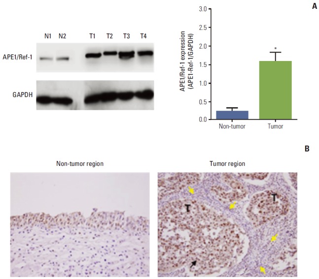
Purpose: Apurinic/apyrimidinic endonuclease 1/redox factor-1 (APE1/Ref-1) is a multifunctional protein that shows elevated expression in a number of cancers. We attempted to determine whether serum APE1/Ref-1 is elevated in patients with bladder cancer.

Materials and methods: Serum APE1/Ref-1 levels were determined using enzyme-linked immunosorbent assay in serum from patients with bladder cancer who had not received chemotherapy or radiotherapy (n=51) and non-tumor controls (n=55). The area under the receiver operating characteristic area under the curve was applied to determine the correlation between clinical factors and the serum levels of APE1/Ref-1.

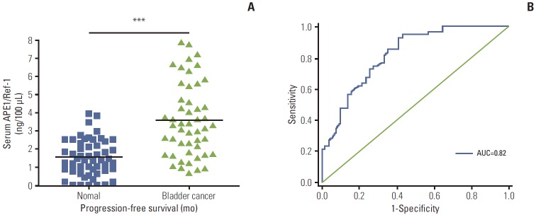
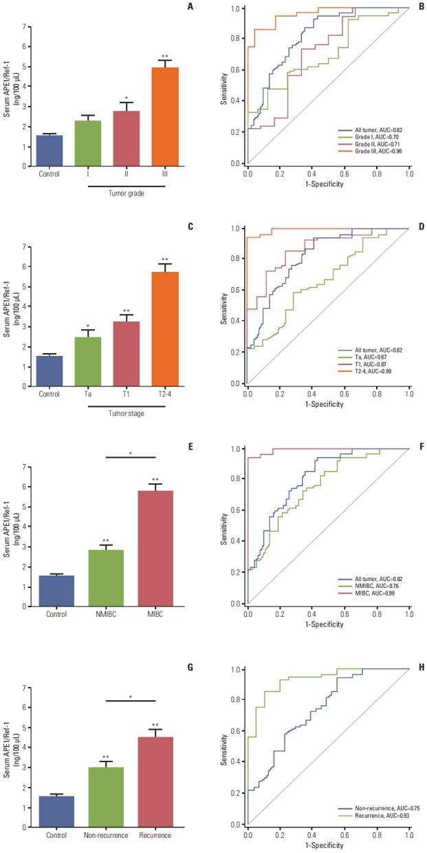
Results: Serum levels of APE1/Ref-1 in bladder cancer patients were significantly elevated compared to those of the control group (3.548 ± 0.333 ng/100 μL [n=51] for bladder cancer vs. 1.547 ± 0.319 ng/100 μL [n=55] for the control group), with a sensitivity and specificity of 93% and 59%, respectively. Serum APE1/Ref-1 levels are associated with tumor stage, grade, muscle invasion, and recurrence.

Conclusion: Serum APE1/Ref-1 might be useful as a potential serologic biomarker for bladder cancer.

Keywords: Apurinic/apyrimidinic endonuclease 1/redox factor-1; Biological markers; Enzyme-linked immunosorbent assay; Urinary bladder neoplasms.


Conflict of interest statement

Conflict of interest relevant to this article was not reported.

Figures

Fig. 1.
Fig. 1.
Serum apurinic/apyrimidinic endonuclease 1/redox factor-1 (APE1/Ref-1) expression is elevated in bladder cancer. (A) Serum APE1/Ref-1 was assayed using an enzyme-linked immunosorbent assay. The results are presented as a scatter plot. Each dot represents one patient (n=55 for non-cancer controls, n=51 for bladder cancer). ***p < 0.01 (compared with the control group). (B) Receiver operating curves of APE1/Ref-1 in bladder cancer detection. The area under curve (AUC) for detection of all cancer by APE1/Ref-1 was 0.82.
Fig. 2.
Fig. 2.
Serum apurinic/apyrimidinic endonuclease1/redox factor-1 (APE1/Ref-1) levels are associated with bladder tumor grade, stage, muscle invasion, and recurrence. Serum APE1/Ref-1 levels were assayed using an enzyme-linked immunosorbent assay. (A) Each bar shows the mean±standard error (SE) (n=16 for grade I, n=12 for grade II, n=23 for grade III, and n=55 for non-cancer controls). *p < 0.05 vs. control, **p < 0.01 vs. control. (B) Receiver operating curves for APE1/Ref-1 detection of different bladder tumor grades. (C) Serum APE1/Ref-1 levels are elevated in patients with higher stage tumors. Each bar shows the mean±SE (n=21 for stage Ta, n=17 for stage T1, n=13 for stage T2-3, and n=55 for non-cancer controls). *p < 0.05 vs. control, **p < 0.01 vs. control. (D) Receiver operating curves for APE1/Ref-1 detection of different bladder tumor stages. (E) Serum APE1/Ref-1 levels are higher in patients with muscle invasive bladder cancer. Each bar shows the mean±SE (n=38 for nonmuscle invasive bladder cancer [NMIBC], n=13 for muscle invasive bladder cancer [MIBC], and n=55 for non-cancer controls). *p < 0.05, **p < 0.01 vs. control. (F) Receiver operating curves for APE1/Ref-1 detection of NMIBC and MIBC. (G) Serum APE1/Ref-1 levels are higher in patients with recurrent tumors. Each bar shows the mean±SE (n=31 for non-recurrence, n=20 for recurrence, and n=55 for non-cancer controls). *p < 0.05, **p < 0.01 vs. control. (H) Receiver operating curves for APE1/Ref-1 detection of recurrent bladder tumors.
Fig. 3.
Fig. 3.
Apurinic/apyrimidinic endonuclease1/redox factor-1 (APE1/Ref-1) expression is elevated in bladder cancer. (A) Representative immunoblot for APE1/Ref-1 in bladder tissues of bladder cancer patients. N, non-tumor tissue as normal tissue; T, tumor tissue. Glyceraldehyde-3-phosphate dehydrogenase (GAPDH) was used as a loading control. Each bar shows the mean±standard error (n=2 for non-tumor, n=4 for tumor tissues). *p < 0.05 vs. non-tumor regions. (B) Immunohistochemical staining for APE1/Ref-1 in bladder cancer. Positive cells are stained brown (black arrow). APE1/Ref-1 expression was clearly elevated in the tumor region, compared with non-tumor regions of bladder tissues. Mononuclear cells were infiltrated close to tumor regions (yellow arrows) (✕400). T, tumor cells.

References

    1. Siegel R, DeSantis C, Virgo K, Stein K, Mariotto A, Smith T, et al. Cancer treatment and survivorship statistics, 2012. CA Cancer J Clin. 2012;62:220–41. - PubMed
    1. Jung KW, Park S, Kong HJ, Won YJ, Lee JY, Park EC, et al. Cancer statistics in Korea: incidence, mortality, survival, and prevalence in 2008. Cancer Res Treat. 2011;43:1–11. - PMC - PubMed
    1. Lamm DL, Blumenstein BA, Crissman JD, Montie JE, Gottesman JE, Lowe BA, et al. Maintenance bacillus Calmette-Guerin immunotherapy for recurrent TA, T1 and carcinoma in situ transitional cell carcinoma of the bladder: a randomized Southwest Oncology Group Study. J Urol. 2000;163:1124–9. - PubMed
    1. Vrooman OP, Witjes JA. Molecular markers for detection, surveillance and prognostication of bladder cancer. Int J Urol. 2009;16:234–43. - PubMed
    1. Raab SS, Grzybicki DM, Vrbin CM, Geisinger KR. Urine cytology discrepancies: frequency, causes, and outcomes. Am J Clin Pathol. 2007;127:946–53. - PubMed

Publication types

Substances