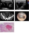
Multiple hemangiomas of the urinary bladder in a child with gross hematuria
- PMID: 25672772
- PMCID: PMC4484289
- DOI: 10.14366/usg.14056
Multiple hemangiomas of the urinary bladder in a child with gross hematuria
Abstract
We report a case of multiple hemangiomas involving the urinary bladder in a 4-year-old boy who presented with recurrent episodes of gross hematuria. On ultrasonography, compared with the bladder wall, the lesions presented as multiple isoechoic polypoid intraluminal masses with mildly increased vascularity on color Doppler exam. Cavernous hemangioma was confirmed by cold-cup biopsy, and the all lesions were coagulated with a Holmium laser. Despite their rarity, bladder hemangiomas should be included in the differential diagnosis of multiple intravesical masses in children with gross hematuria.
Keywords: Child, preschool; Hemangioma; Ultrasonography; Urinary bladder.
Conflict of interest statement
No potential conflict of interest relevant to this article was reported.
Figures

References
-
- Greenfield SP, Williot P, Kaplan D. Gross hematuria in children: a ten-year review. Urology. 2007;69:166–169. - PubMed
-
- Huppmann AR, Pawel BR. Polyps and masses of the pediatric urinary bladder: a 21-year pathology review. Pediatr Dev Pathol. 2011;14:438–444. - PubMed
-
- Bates DG. Bladder and urethra. In: Coley BD, editor. Caffey's pediatric diagnostic imaging. 12th ed. Philadelphia: Saunders; 2013. pp. 1262–1273.
-
- Numanoglu KV, Tatli D. A rare cause of hemorrhagic shock in children: bladder hemangioma. J Pediatr Surg. 2008;43:e1–e3. - PubMed
-
- Kogan MG, Koenigsberg M, Laor E, Bennett B. US case of the day. Cavernous hemangioma of the bladder. Radiographics. 1996;16:443–447. - PubMed
Publication types
LinkOut - more resources
Full Text Sources
Other Literature Sources

