. 2015 Jun;11(6):4079-86.
doi: 10.3892/mmr.2015.3339. Epub 2015 Feb 12.

Association between oxidative DNA damage and the expression of 8-oxoguanine DNA glycosylase 1 in lung epithelial cells of neonatal rats exposed to hyperoxia

Affiliations

Association between oxidative DNA damage and the expression of 8-oxoguanine DNA glycosylase 1 in lung epithelial cells of neonatal rats exposed to hyperoxia

Linlin Jin et al. Mol Med Rep. 2015 Jun.

Abstract

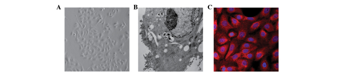
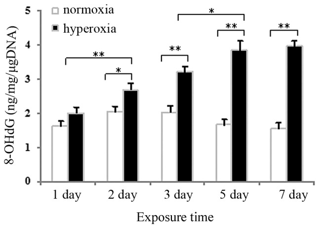
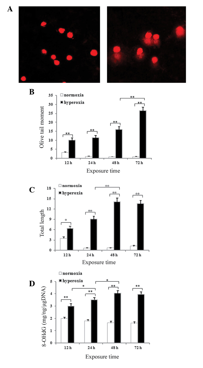
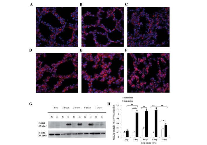
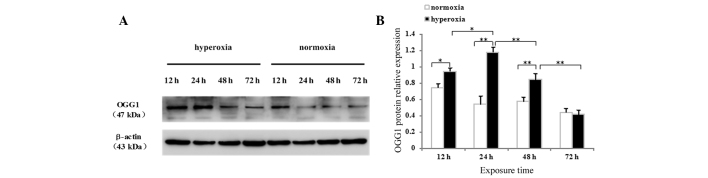
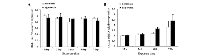
Previous studies have demonstrated that oxidative stress‑induced lung injury is involved in the occurrence and developmental process of bronchopulmonary dysplasia (BPD). The present study assessed whether oxidative DNA damage occurs in the early stages of hyperoxia‑induced BPD in neonatal rats and evaluated the expression and localization of the DNA repair gene, 8‑oxoguanine DNA glycosylase 1 (OGG1), upon exposure to hyperoxia. Neonatal rats and primary cultured neonatal rat alveolar epithelial type II (AECII) cells were exposed to hyperoxia (90% O2) or normoxia (21% O2) and the expression levels of 8‑hydroxy‑2'‑deoxyguanosine (8‑OHdG) in the lung tissues and AECII cells were determined using a competitive enzyme‑linked immunosorbent assay. DNA strand breaks in the AECII cells were detected using a comet assay. The expression and localization of the OGG1 protein in the lung tissues and AECII cells were determined by immunofluorescence confocal microscopy and western blotting. The mRNA expression levels of OGG1 in the lung tissues and AECII cells were determined by reverse transcription polymerase chain reaction. The expression of 8‑OHdG was elevated in the hyperoxia‑exposed neonatal rat lung tissue and the AECII cells compared with the normoxic controls. The occurrence of DNA strand breaks in the AECII cells increased with increasing duration of hyperoxia exposure. The protein expression of OGG1 was significantly increased in the hyperoxia‑exposed lung tissues and AECII cells, with OGG1 preferentially localized to the cytoplasm. No concomitant increase in the mRNA expression of OGG1 was detected. These results revealed that oxidative DNA damage occurred in lung epithelial cells during early‑stage BPD, as confirmed by in vitro and in vivo hyperoxia exposure experiments, and the increased expression of OGG1 was associated with this process.


Figures

Figure 1
Figure 1
Identification of cultured neonatal rat alveolar epithelial type II cells. (A) Inverted phase contrast microscopy (magnification, ×200). (B) Transmission electron microscopy (magnification, ×5,000). (C) Surfactant protein-C detected by immunofluorescence staining (magnification, ×400).
Figure 2
Figure 2
Hyperoxia-induced DNA damage in neonatal rat lung tissues. Competitive enzyme-linked immunosorbent assay was used to detect 8-OHdG in the rat lung tissues. Data are expressed as the mean ± standard deviation of the mean (n=6 per group; *P<0.05; **P<0.01 as compared with the normoxia group). 8-OHdG, 8-hydroxy-2′-deoxyguanosine.
Figure 3
Figure 3
Hyperoxia-induced DNA damage in cultured AECII cells. (A) Fluorescence of ‘comets’ in alveolar epithelial type II cells under normoxia and hyperoxia for 48 h (magnification, ×400), following which the (B) olive tail moments, (C) tail lengths and (D) 8-OHdG DNA were quantified. Data are expressed as the mean ± standard deviation (*P<0.05 and **P<0.01 as compared with the normoxia group). 8-OHdG, 8-hydroxy-2′-deoxyguanosine.
Figure 4
Figure 4
Expression of OGG1 (red stain) is predominantly localized in the cytoplasm on (A) day 1 and (B) day 5 in normoxia-exposed rats and on (C) day 1 in hyperoxia-exposed rats. On days (D) 3, (E) 5 and (F) 7 in hyperoxia (magnification, ×400), the expression of OGG1 increased in the cytoplasm and the nucleus. (G and H) Western blotting revealed similar patterns of expression. Data are expressed as the mean ± standard deviation (*P<0.05, **P<0.01 as compared with the normoxia group). OGG1, 8-oxoguanine DNA glycosylase 1; N, normoxia; H, hyperoxia.
Figure 5
Figure 5
Protein expression of OGG1 in cultured neonatal rat alveolar epithelial type II cells. (A) Western blotting and (B) densitometric quantification of the protein expression of OGG1 following different durations of hyperoxia or normoxia exposure. Data are expressed as the mean ± standard deviation (*P<0.05; **P<0.01 as compared with the normoxia group). OGG1, 8-oxoguanine DNA glycosylase 1.
Figure 6
Figure 6
mRNA expression levels of OGG1 in lung tissues and AECII cells. (A) Neonatal rat lung tissues and (B) neonatal rat AECII cells exposed to hyperoxia or normoxia. Data are expressed as the mean ± standard deviation (P>0.05 for all comparisons). AECII, alveolar epithelial type II cells; OGG1, 8-oxoguanine DNA glycosylase 1.

References

    1. Jobe AH, Bancalari E. Bronchopulmonary dysplasia. Am J Respir Crit Care Med. 2001;163:1723–1729. doi: 10.1164/ajrccm.163.7.2011060. - DOI - PubMed
    1. Bancalari E, Claure N, Sosenko IR. Bronchopulmonary dysplasia: Changes in pathogenesis, epidemiology and definition. Semin Neonatol. 2003;8:63–71. doi: 10.1016/S1084-2756(02)00192-6. - DOI - PubMed
    1. Greenough A, Cox S, Alexander J, et al. Health care utilisation of infants with chronic lung disease, related to hospitalisation for RSV infection. Arch Dis Child. 2001;85:463–468. doi: 10.1136/adc.85.6.463. - DOI - PMC - PubMed
    1. Saigal S, Doyle LW. An overview of mortality and sequelae of preterm birth from infancy to adulthood. Lancet. 2008;371:261–269. doi: 10.1016/S0140-6736(08)60136-1. - DOI - PubMed
    1. Reyburn B, Martin RJ, Prakash YS, et al. Mechanisms of injury to the preterm lung and airway: implications for long-term pulmonary outcome. Neonatology. 2012;101:345–352. doi: 10.1159/000337355. - DOI - PMC - PubMed

Publication types

MeSH terms