Review
. 2014 Nov 13;18(6):557.
doi: 10.1186/s13054-014-0557-2.

Aneurysmal subarachnoid haemorrhage from a neuroimaging perspective

Review

Aneurysmal subarachnoid haemorrhage from a neuroimaging perspective

Airton Leonardo de Oliveira Manoel et al. Crit Care. .

Abstract

Neuroimaging is a key element in the management of patients suffering from subarachnoid haemorrhage (SAH). In this article, we review the current literature to provide a summary of the existing neuroimaging methods available in clinical practice. Noncontrast computed tomography is highly sensitive in detecting subarachnoid blood, especially within 6 hours of haemorrhage. However, lumbar puncture should follow a negative noncontrast computed tomography scan in patients with symptoms suspicious of SAH. Computed tomography angiography is slowly replacing digital subtraction angiography as the first-line technique for the diagnosis and treatment planning of cerebral aneurysms, but digital subtraction angiography is still required in patients with diffuse SAH and negative initial computed tomography angiography. Delayed cerebral ischaemia is a common and serious complication after SAH. The modern concept of delayed cerebral ischaemia monitoring is shifting from modalities that measure vessel diameter to techniques focusing on brain perfusion. Lastly, evolving modalities applied to assess cerebral physiological, functional and cognitive sequelae after SAH, such as functional magnetic resonance imaging or positron emission tomography, are discussed. These new techniques may have the advantage over structural modalities due to their ability to assess brain physiology and function in real time. However, their use remains mainly experimental and the literature supporting their practice is still scarce.


Figures
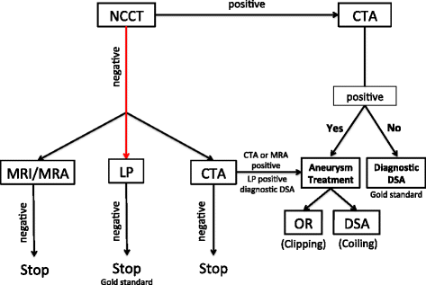
Figure 1
Figure 1
Diagnostic approach for subarachnoid haemorrhage in patients presenting with more than 6 hours of headache onset. CTA, computed tomography angiography; DSA, digital subtraction angiography; LP, lumbar puncture; MRA, magnetic resonance angiography; MRI, magnetic resonance imaging; NCCT, noncontrast computed tomography; OR, operating room.
Figure 2
Figure 2
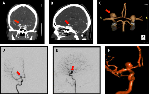
Computed tomography angiography and digital subtraction angiography. (A) to (C) Computed tomography angiography. (D) to (F) Digital subtraction angiography. Red arrow, right terminal internal carotid artery aneurysm. (A), (D) Coronal view. (B), (E) Sagittal view. (C), (F) Three-dimensional rendering.
Figure 3
Figure 3
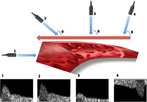
Technical aspects. Transcranial Doppler ultrasonography (TCD) probe emits low-frequency (2 MHz), pulsed waves that cross the skull at specific points called acoustic windows. The emitted waves are reflected back from the moving red blood cells at an altered frequency fe. The Doppler effect (fe) is the difference in frequency between the transmitted wave f0 and the received wave fe: fd = fe – f0. The red blood cells’ velocity can then be calculated according to the following mathematic equation: V = c × fd / 2 × f0 × cosθ. θ is the angle formed by the ultrasound beam and the blood flow. Ideally this angle should approach 0° so that the ultrasound beam is parallel to the blood flow (cos0° = 1). The higher this angle, the lower the velocity read by the machine. If the ultrasound beam is perpendicular to the blood flow, no velocity will be detected (cos90° = 0). This concept is fundamental to the performance and interpretation of TCD results. The velocities can be underestimated, never overestimated.
Figure 4
Figure 4
Effect of the angle between the ultrasound beam and blood flow. (1) Ultrasound beam is parallel to blood flow, acquiring an optimal, high-frequency Doppler signal. (2) Ultrasound beam forms an angle <20°, leading to a good signal. (3) Ultrasound beam is almost at 90°, resulting in a damped waveform. (4) Blood flow direction is away from ultrasound beam, which results in a negative Doppler signal.
Figure 5
Figure 5
Multimodal computed tomography. (A) Computed tomography (CT) angiography (coronal plane) shows severe angiographic vasospasm in the left middle cerebral artery (MCA; red arrow). (B) CT perfusion displays increased mean transit time (increased shades of red in the left hemisphere) due to MCA vasospasm. (C) Same patient, CT perfusion repeated after 48 hours of haemodynamic augmentation, which shows resolution of perfusion changes. (D) Noncontrast computed tomography (NCCT) showing the measurement of bicaudate index (A/B) as proposed by van Gijn and colleagues [98]. (E) NCCT shows large volume of intraventricular haemorrhage. The tip of the bilateral external ventriculostomy drains can be seen in the anterior horn of the lateral ventricles (blue arrows). (F) NCCT quality severely compromised by artefacts generated by the metallic colis.
Figure 6
Figure 6
Digital subtraction angiography. (A) Selective left vertebral artery injection shows a basilar tip aneurysm (red arrow). (B) The same basilar tip aneurysm (white arrow), now appreciated through a three-dimensional rendering. (C) The gray structure is the mass of coils packing the previous basilar tip aneurysm. Final results show a complete obliteration of the aneurysm’s sac after the endovascular coiling.
Figure 7
Figure 7
Magnetic resonance angiography. (A) Contrast-enhanced (CE) magnetic resonance angiography (MRA) sequence showing recanalisation of a previously coiled basilar tip aneurysm (large white arrow). (B) Time-of-flight MRA sequence revealing the refilling of the same basilar tip aneurysm (small white arrow). (C) CE-MRA and (D) time-of-flight MRA sequences after retreatment. No additional filling of the aneurysm is seen (red arrows).

References

    1. Shea BJ, Grimshaw JM, Wells GA, Boers M, Andersson N, Hamel C, Porter AC, Tugwell P, Moher D, Bouter LM. Development of AMSTAR: a measurement tool to assess the methodological quality of systematic reviews. BMC Med Res Methodol. 2007;7:10. doi: 10.1186/1471-2288-7-10. - DOI - PMC - PubMed
    1. Diringer MN, Bleck TP, Claude Hemphill J, 3rd, Menon D, Shutter L, Vespa P, Bruder N, Connolly ES, Jr, Citerio G, Gress D, Hänggi D, Hoh BL, Lanzino G, Le Roux P, Rabinstein A, Schmutzhard E, Stocchetti N, Suarez JI, Treggiari M, Tseng MY, Vergouwen MD, Wolf S, Zipfel G, Neurocritical Care Society Critical care management of patients following aneurysmal subarachnoid hemorrhage: recommendations from the Neurocritical Care Society’s multidisciplinary consensus conference. Neurocrit Care. 2011;15:211–240. doi: 10.1007/s12028-011-9605-9. - DOI - PubMed
    1. Connolly ES, Jr, Rabinstein AA, Carhuapoma JR, Derdeyn CP, Dion J, Higashida RT, Hoh BL, Kirkness CJ, Naidech AM, Ogilvy CS, Patel AB, Thompson BG, Vespa P. Guidelines for the management of aneurysmal subarachnoid hemorrhage: a guideline for healthcare professionals from the American Heart Association/American Stroke Association. Stroke. 2012;43:1711–1737. doi: 10.1161/STR.0b013e3182587839. - DOI - PubMed
    1. Steiner T, Juvela S, Unterberg A, Jung C, Forsting M, Rinkel G. European Stroke Organization guidelines for the management of intracranial aneurysms and subarachnoid haemorrhage. Cerebrovasc Dis. 2013;35:93–112. doi: 10.1159/000346087. - DOI - PubMed
    1. Edlow JA, Panagos PD, Godwin SA, Thomas TL, Decker WW. Clinical policy: critical issues in the evaluation and management of adult patients presenting to the emergency department with acute headache. Ann Emerg Med. 2008;52:407–436. doi: 10.1016/j.annemergmed.2008.07.001. - DOI - PubMed

Publication types