. 2015 Apr;96(4):313-23.
doi: 10.1007/s00223-015-9954-z. Epub 2015 Feb 13.

High-fat diet causes bone loss in young mice by promoting osteoclastogenesis through alteration of the bone marrow environment

Affiliations

High-fat diet causes bone loss in young mice by promoting osteoclastogenesis through alteration of the bone marrow environment

Lei Shu et al. Calcif Tissue Int. 2015 Apr.

Abstract

Obesity is a severe health problem in children, afflicting several organ systems including bone. However, the role of obesity on bone homeostasis and bone cell function in children has not been studied in detail. Here we used young mice fed a high-fat diet (HFD) to model childhood obesity and investigate the effect of HFD on the phenotype of cells within the bone marrow environment. Five-week-old male mice were fed a HFD for 3, 6, and 12 weeks. Decreased bone volume was detected after 3 weeks of HFD treatment. After 6 and 12 weeks, HFD-exposed mice had less bone mass and increased osteoclast numbers. Bone marrow cells, but not spleen cells, from HFD-fed mice had increased osteoclast precursor frequency, elevated osteoclast formation, and bone resorption activity, as well as increased expression of osteoclastogenic regulators including RANKL, TNF, and PPAR-gamma. Bone formation rate and osteoblast and adipocyte numbers were also increased in HFD-fed mice. Isolated bone marrow cells also had a corresponding elevation in the expression of positive regulators of osteoblast and adipocyte differentiation. Our findings indicate that in juvenile mice, HFD-induced bone loss is mainly due to increased osteoclast bone resorption by affecting the bone marrow microenvironment. Thus, targeting osteoclast formation may present a new therapeutic approach for bone complications in obese children.


Figures
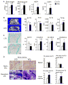
Figure 1
Figure 1. Decreased bone volume and increased osteoclasts in mice fed a HFD for 12 weeks
Five-week-old male mice were fed a high-fat diet (HFD) or lean diet (LFD) for 12 weeks. (A) Body weight, blood glucose, serum leptin, and total non-esterified fatty acids (NEFA) were measured. (B) Representative μCT scans (bar = 100 μm) and morphometric data of BV/TV, trabecular number (Tb.N), trabecular separation (Tb.Sp) and trabecular thickness (Tb.Th) in the femoral bone. (C) TRAP-stained femoral sections (bar = 250 μm) show decreased trabecular bone volume and increased osteoclasts in HFD-fed mice. Histomorphometric analyses including bone volume, adipocyte and osteoclast parameters were performed. (D) Spleen cells or bone marrow cells were cultured with M-CSF and RANKL for 5–7 days on plastic for osteoclast formation or for 10–12 days on bone slides for bone resorption. The area of TRAP+ osteoclasts and resorption pits were counted. Values are mean ± SD of 6 mice or 5 wells. *, p<0.05; **, p<0.01, ***, p<0.001 versus lean group.
Figure 2
Figure 2. Increased osteoblast function in mice fed a HFD
Mice that were fed a HFD for 12 weeks were used. (A) Calcein labeled trabecular and cortical bones of femurs from mice fed with LFD and HFD. Mineral apposition rate (MAR), bone formation rate (BFR), and mineral surface per bone surface (MS/BS) were calculated. Values are mean ± SD of 4 mice. (B) Left panel: expression levels of osteoblast-related genes in CD45 cells isolated from bone marrow cells. Right panels: osteoblast differentiation (Alkaline phosphatase staining) and mineralization (Alizarin Red staining) were determined in bone marrow stromal cells after they were cultured in the osteoblast inducing medium for 14 and 21 days, respectively. (C) Left panel: expression levels of adipogenic genes in CD45 cells isolated from bone marrow cells. Right panels: adipocyte differentiation was assessed in cells that were cultured with the adipocyte-induced medium for 7 days. *, p<0.05; **, p<0.01, ***, p<0.001, versus lean mice.
Figure 3
Figure 3. Decreased bone volume and increased osteoclasts in mice fed short-term HFD
Five-week-old male mice were fed a HFD or LFD for 6 weeks. (A) Body weight and blood glucose levels were measured. (B) Representative μCT scans (bar = 100 μm) and morphometric data of BV/TV, Tb.N, Tb.Sp and Tb.Th in the vertebral bone. (C) TRAP-stained femoral sections (bar = 250 μm) show decreased trabecular bone volume and increased osteoclasts in HFD-fed mice. Histomorphometry including bone volume, adipocyte and osteoclast parameters were measured. (D) Bone marrow cells were cultured with M-CSF and RANKL for 5–7 days on plastic for osteoclast formation. The area of TRAP+ osteoclasts was counted. Values are mean ± SD of 6 mice or 5 wells. *, p<0.05; **, p<0.01, ***, p<0.001 versus lean group.
Figure 4
Figure 4. Increased osteoclast precursors in cells from bone marrow, but not from the spleen, of mice fed a HFD
Mice that were fed a HFD for 6 weeks were used. (A) Osteoclast precursors defined as c-kit+, c-fms+ or CD11b+ cells were examined by FACS analysis. The percentage (B) and total number (C) of c-kit+, c-fms+ or CD11b+ cells vs total of bone marrow and spleen mononuclear cells were assessed. Values are mean ± SD of 3 mice per group. (D) Bone marrow cells were cultured with a series dilution of M-CSF and RANKL for 5–7 days to generate osteoclasts. The area of TRAP+ osteoclasts was counted. Values are mean ± SD of 5 wells. *, p<0.05; **, p<0.01, ***, p<0.001, versus cells from lean group.
Figure 5
Figure 5. Alteration of osteoclastogenic genes in bone marrow cells of mice fed a HFD
Mice that were fed a HFD for 6 weeks were used. CD45 and CD45+ cells were isolated from bone marrow or spleen using micro-beads conjugated with anti-mouse CD45 antibody. Relative expression levels of genes of interest were determined by qPCR. (A) The fold increases of osteoclastogenic gene expression in CD45 and CD45+ cells that were isolated from bone marrow and spleen. (B) The fold changes of adipogenic gene expression in CD45+ cells that were isolated from bone marrow and spleen. Values are mean ± SD of 3 mice per group. *, p<0.05; **, p<0.01, versus lean mice.
Figure 6
Figure 6. A model of HFD induced bone loss in young mice
In the bone marrow cavity of young mice, HFD affects both CD45 mesenchymal cells and CD45+ hematopoietic cells. HFD promotes osteoblastogenesis and adipogenesis by increasing the expression of positive regulators of osteoblasts and adipocytes in osteoprogenitors. At the same time, HFD stimulates osteoclast formation by up-regulating expression of osteoclastogenic factors, which increases the formation and differentiation of osteoclast precursors. Increased osteoclastic bone resorption overrides increased osteoblastic bone formation, resulting in net bone loss.

References

    1. Dimitri P, Bishop N, Walsh JS, Eastell R. Obesity is a risk factor for fracture in children but is protective against fracture in adults: a paradox. Bone. 2012;50:457–466. - PubMed
    1. Looker AC, Flegal KM, Melton LJ., 3rd Impact of increased overweight on the projected prevalence of osteoporosis in older women. Osteoporos Int. 2007;18:307–313. - PubMed
    1. Albala C, Yanez M, Devoto E, Sostin C, Zeballos L, Santos JL. Obesity as a protective factor for postmenopausal osteoporosis. Int J Obes Relat Metab Disord. 1996;20:1027–1032. - PubMed
    1. Cao JJ, Gregoire BR, Gao H. High-fat diet decreases cancellous bone mass but has no effect on cortical bone mass in the tibia in mice. Bone. 2009;44:1097–1104. - PubMed
    1. Rana AR, Michalsky MP, Teich S, Groner JI, Caniano DA, Schuster DP. Childhood obesity: a risk factor for injuries observed at a level-1 trauma center. J Pediatr Surg. 2009;44:1601–1605. - PMC - PubMed

Publication types

MeSH terms

LinkOut - more resources