. 2015 May;89(9):4738-47.
doi: 10.1128/JVI.02296-14. Epub 2015 Feb 11.

The glycosylation status of PrPC is a key factor in determining transmissible spongiform encephalopathy transmission between species

Affiliations

The glycosylation status of PrPC is a key factor in determining transmissible spongiform encephalopathy transmission between species

Frances K Wiseman et al. J Virol. 2015 May.

Abstract

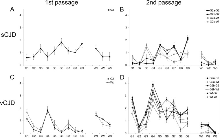
The risk of transmission of transmissible spongiform encephalopathies (TSE) between different species has been notoriously unpredictable because the mechanisms of transmission are not fully understood. A transmission barrier between species often prevents infection of a new host with a TSE agent. Nonetheless, some TSE agents are able to cross this barrier and infect new species, with devastating consequences. The host PrP(C) misfolds during disease pathogenesis and has a major role in controlling the transmission of agents between species, but sequence compatibility between host and agent PrP(C) does not fully explain host susceptibility. PrP(C) is posttranslationally modified by the addition of glycan moieties which have an important role in the infectious process. Here, we show in vivo that glycosylation of the host PrP(C) has a significant impact on the transmission of TSE between different host species. We infected mice carrying different glycosylated forms of PrP(C) with two human agents (sCJDMM2 and vCJD) and one hamster strain (263K). The absence of glycosylation at both or the first PrP(C) glycosylation site in the host results in almost complete resistance to disease. The absence of the second site of N-glycan has a dramatic effect on the barrier to transmission between host species, facilitating the transmission of sCJDMM2 to a host normally resistant to this agent. These results highlight glycosylation of PrP(C) as a key factor in determining the transmission efficiency of TSEs between different species.

Importance: The risks of transmission of TSE between different species are difficult to predict due to a lack of knowledge over the mechanisms of disease transmission; some strains of TSE are able to cross a species barrier, while others do not. The host protein, PrP(C), plays a major role in disease transmission. PrP(C) undergoes posttranslational glycosylation, and the addition of these glycans may play a role in disease transmission. We infected mice that express different forms of glycosylated PrP(C) with three different TSE agents. We demonstrate that changing the glycosylation status of the host can have profound effects on disease transmission, changing host susceptibility and incubation times. Our results show that PrP(C) glycosylation is a key factor in determining risks of TSE transmission between species.


Figures

FIG 1
FIG 1
Lesion profile analysis of wild-type (Wt) and G2 mice after intracerebral inoculation with sCJDMM2 (A), second passage of sCJDMM2 (B), vCJD (C), and second passage of vCJD (D). The second passage was carried out from selected G2 and wild-type mice showing TSE vacuolation and/or PrP. Group size, n ≥ 6 (± standard errors of the means). Gray matter scoring regions are labeled G1 to G9: G1, dorsal medulla; G2, cerebellar cortex; G3, superior colliculus; G4, hypothalamus; G5, thalamus; G6, hippocampus; G7, septum; G8, retrosplenial cortex; G9, cingulate and adjacent motor cortex. White-matter scoring regions are labeled W1 to W3: W1, cerebellar white matter; W2, mesencephalic tegmentum; W3, pyramidal tract.
FIG 2
FIG 2
PrP deposition in the brains of wild-type and G2 mice after intracerebral inoculation with sCJDMM2, vCJD, or 263K agent. (A) G2 mouse inoculated with sCJDMM2; (B) G2 mouse inoculated with vCJD; (C) G2 mouse inoculated with 263K; (D) wild-type mouse inoculated with sCJDMM2; (E) wild-type mouse inoculated with vCJD; (F) wild-type mouse inoculated with 263K. Arrows indicate examples of PrP accumulation in the form of plaque-like deposits in panels A and B and examples of fine punctate PrP accumulation in panels C, E, and F. No PrP accumulation was detected in panel D. PrP was detected with 6H4 antibody. dg, dentate gyrus; cc, corpus callosum. Scale bars, 500 μm.
FIG 3
FIG 3
Biochemical analysis of PrPSc from the brains of G2 and wild-type (Wt) mice inoculated with sCJDMM2, vCJD, and 263K. (A) PrPSc was isolated from four sCJD-challenged G2 mice, one vCJD-challenged G2 mouse (assayed in duplicate), and one vCJD-challenged wild-type mouse (assayed in duplicate) by standard PK digestion. PrPSc was concentrated by NaPTA precipitation. The amount of PrPSc in the vCJD-challenged G2 sample was too low to detect. Uninfected mice and a 263K-challenged wild-type mouse were included as controls. (B) PrPSc was isolated from vCJD-challenged G2 mice by standard PK digestion followed by NaPTA precipitation. The amount of PrPSc was equalized by dilution following NaPTA precipitation to allow comparison (equivalent of 5 μg of G2 brain and 0.5 to 1 μg of wild-type brain). Uninfected mice were included as controls. (C) PrPSc was isolated from three 263K-challenged G2 mice and three wild-type mice by standard PK digestion. Uninfected mice and a 263K-challenged hamster were included as controls. The different isoforms of PrP are denoted Di (for diglycosylated), Mono (monoglycosylated), and Un (unglycosylated). PrPSc was detected with 8H4.
FIG 4
FIG 4
PrPC expression levels in the brains of glycosylation-deficient transgenic mice. Representative Western blots showing the different PrPC isoforms in wild-type (Wt), G1, G2, and G3 mice using BC6 (A) and BH1 (B) antibodies. Western blots underwent densitometry to measure levels of PrPC. α-Tubulin was used as a loading control. The different isoforms of PrPC are denoted Di (for diglycosylated), Mono (monoglycosylated), Un (unglycosylated), and C1 frag. (C1 fragments).

References

    1. Prusiner SB. 1998. Prions. Proc Natl Acad Sci U S A 95:13363–13383. doi:10.1073/pnas.95.23.13363. - DOI - PMC - PubMed
    1. Will RG. 2003. Acquired prion disease: iatrogenic CJD, variant CJD, kuru. Br Med Bull 66:255–265. doi:10.1093/bmb/66.1.255. - DOI - PubMed
    1. Bueler H, Aguzzi A, Sailer A, Greiner RA, Autenried P, Aguet M, Weissmann C. 1993. Mice devoid of PrP are resistant to scrapie. Cell 73:1339–1347. doi:10.1016/0092-8674(93)90360-3. - DOI - PubMed
    1. Manson JC, Clarke AR, Hooper ML, Aitchison L, McConnell I, Hope J. 1994. 129/Ola mice carrying a null mutation in PrP that abolishes mRNA production are developmentally normal. Mol Neurobiol 8:121–127. doi:10.1007/BF02780662. - DOI - PubMed
    1. Prusiner SB. 1982. Novel proteinaceous infectious particles cause scrapie. Science 216:136–144. doi:10.1126/science.6801762. - DOI - PubMed

Publication types