. 2015 Apr 1;26(7):1323-44.
doi: 10.1091/mbc.E14-11-1531. Epub 2015 Feb 11.

Cytosolic chaperones mediate quality control of higher-order septin assembly in budding yeast

Affiliations

Cytosolic chaperones mediate quality control of higher-order septin assembly in budding yeast

Courtney R Johnson et al. Mol Biol Cell. .

Abstract

Septin hetero-oligomers polymerize into cytoskeletal filaments with essential functions in many eukaryotic cell types. Mutations within the oligomerization interface that encompasses the GTP-binding pocket of a septin (its "G interface") cause thermoinstability of yeast septin hetero-oligomer assembly, and human disease. When coexpressed with its wild-type counterpart, a G interface mutant is excluded from septin filaments, even at moderate temperatures. We show that this quality control mechanism is specific to G interface mutants, operates during de novo septin hetero-oligomer assembly, and requires specific cytosolic chaperones. Chaperone overexpression lowers the temperature permissive for proliferation of cells expressing a G interface mutant as the sole source of a given septin. Mutations that perturb the septin G interface retard release from these chaperones, imposing a kinetic delay on the availability of nascent septin molecules for higher-order assembly. Un-expectedly, the disaggregase Hsp104 contributes to this delay in a manner that does not require its "unfoldase" activity, indicating a latent "holdase" activity toward mutant septins. These findings provide new roles for chaperone-mediated kinetic partitioning of non-native proteins and may help explain the etiology of septin-linked human diseases.


Figures

FIGURE 1:
FIGURE 1:
Alternative alleles of a given septin subunit compete to occupy a limiting number of positions within hetero-octamers. (A) “Quality control” of higher-order septin assembly in budding yeast. Left, schematic illustration of the localization of a GFP-tagged wild-type (CDC10+) or mutant (cdc10(D182N)-GFP) septin protein after introduction of the plasmid-encoded gene into wild-type (CDC10+/CDC10+) diploid cells. Middle, transmitted light and GFP fluorescence (inverted grayscale) micrographs of BY4743 cells carrying the cdc10(D182N)-GFP plasmid pCdc10-1-GFP, grown to mid log phase at 22°C. Right, schematic illustration of line scans of fluorescence micrographs, with actual data from individual cells. An eight-pixel-wide line was drawn perpendicular to the axis of the septin ring and used to plot a profile of fluorescence signal. The height of the peak (for neck localization) or depth of the trough (for neck exclusion) was calculated as shown. (B–E) Bud neck fluorescence for the indicated plasmid-encoded, fluorescently tagged mutant septin (bracketed genotype) expressed in cells of the indicated chromosomal genotype (genotype without brackets). Error bars, mean with SEM; n, number of cells analyzed. Septin hetero-octamers are illustrated below as in A, with multiple polypeptides competing for occupancy of the same position shown horizontally adjacent to each other. Leftmost, septin subunits identified by numbers: 1, Shs1; 3, Cdc3; 10, Cdc10; 11, Cdc11; 12, Cdc12. Fluorescently tagged subunits are solid green, absent subunits are indicated with dashed lines, and asterisks and daggers indicate G interface/NBP mutations. Shs1 is shown in blue. Strains: BY4743 (CDC10+/CDC10+), BY4741 (CDC12+), CBY07236 (cdc3(G365R)), CBY06417 (cdc10(D182N)), CBY08756 (cdc11(G32E)), CBY05110 (cdc12(G247E)), and JTY3631 (shs1∆). Plasmids: pLA10 ([CDC10-GFP]); pCdc10-1-GFP ([cdc10(D182N)-GFP]); pML51 ([cdc11(K172A D174A)-YFP]); pML114, which encodes Cdc12(G44V K47E T48N)-YFP ([cdc12*-YFP]); and YCpH-cdc12-1-GFP, which encodes Cdc12(G247E)-GFP ([G247E-GFP]). (F) Quantitative immunoblotting to compare levels of Cdc10-GFP expressed from the endogenous CDC10 locus was performed with whole-cell protein extracts of CDC10-GFP strains carrying the wild-type (CDC12+) or cdc12(G247E) allele at the CDC12 locus. After separation of proteins by 4–20% gradient SDS–PAGE and transfer to PVDF, immunoblot analysis was performed using antibodies recognizing GFP and appropriate fluorescently labeled secondary antibodies (top blot). Right, molecular weights of the Li-Cor Chameleon Duo Pre-stained Protein Ladder (928-60000; Li-Cor) indicated with arrows. After scanning and quantifying the GFP signal, the membrane was exposed to antibodies recognizing the loading control Zwf1 (glucose-6-phosphate dehydrogenase) and appropriate secondary antibodies (bottom blot). Left, arrows and labels indicate Cdc10-GFP and Zwf1; gray arrow, Cdc10-GFP signal detected in the Zwf1 scan. Signal for each band was quantified by subtracting the background signal from an equivalent area from a signal-free part of the same lane, then dividing the Cdc10-GFP signal by the Zwf1 signal; each of these values was normalized to this value for the first CDC12+ sample and plotted below. CDC12+ strains were MMY0166 and MMY0167, and cdc12(G247E) strains were MMY0168, MMY0169, and MMY0170.
FIGURE 2:
FIGURE 2:
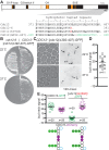
Mutant septins with native G interfaces evade exclusion by quality control. (A) Top, schematic layout of Cdc12 protein domains/motifs, including major elements of the GTP-binding domain (G1/P loop, G3/switch II, and G4) and septin–septin interfaces (G3/switch II, septin-unique element [SUE]) and highlighting the hydrophobic heptad repeats within the extreme C- terminus predicted to form a coiled coil (c.c.). Orange bars, approximate locations of homologous residues buried in the human septin G dimer interface (Sirajuddin et al., 2007, 2009). Bottom, primary sequence of the c.c. region, illustrating the mutations in the Cdc12-6, Cdc12(K391N), and Cdc12(∆392-407)-GFP proteins. (B) Cells of the indicated genotype (plasmid-encoded genes are indicated with brackets) carrying a plasmid (YCpcdc12-6-GFP) encoding Cdc12(∆392-407)-GFP were streaked on solid medium selective for the plasmid and incubated at 22 or 37°C for 3 d. Strains were YEF473A (CDC12+) and M-1726 (cdc12-6). (C) As in Figure 1A, transmitted light and fluorescence micrographs of CDC12+ cells (strain YEF473a) carrying plasmid YCpcdc12-6-GFP grown to mid log phase in selective medium at the indicated temperatures. Arrowheads, fluorescence signal enriched at the bud neck. As indicated, 120-ms exposures were taken to prevent signal saturation. (D) As in Figure 1, B–E, for wild-type strain BY4741 and plasmid YCpcdc12-6-GFP (∆392-407) grown at 22°C. (E) As in Figure 1, B–E, for the CDC12+ CDC10-mCherry strain JTY3992 and plasmid pMVB62 ([CDC12-GFP]) or YEpLcdc12(G268R)-GFP ([cdc12(G268R)-GFP]). Purple, the mCherry tag on Cdc10, fluorescence of which was also quantified as a control for bud neck signal.
FIGURE 3:
FIGURE 3:
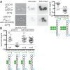
Exclusion of mutant septins by quality control acts before G dimerization and cannot be overcome by mutant overexpression. (A) Left, as in Figure 1A, but with haploid CDC10+ cells (strain BY4742) and plasmids with which Cdc10-GFP (pMETp-Cdc10-GFP) or Cdc10(D182N)-GFP (pPmet-Cdc10-1-GFP) is expressed from the high-level MET17 promoter in growth medium lacking methionine. The 60- and 250-ms exposures are shown for comparison to micrographs in other figures, which always used 250-ms exposures unless specified otherwise. Right, quantification of bud neck signals and illustration of septin subunit composition. (B) As in A, right, with plasmid pLA10K ([CDC10-GFP]) or YCpK-Cdc10-1-GFP ([cdc10*-GFP*]) and cdc3(D210G) strain MMY0131 (CDC10+) or MMY0130 (cdc10(D182N)).
FIGURE 4:
FIGURE 4:
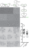
Mutant septins evade exclusion by quality control if assembled into hetero-octamers before introduction of the wild-type allele. Top, fusion of the SNAP-Tag (SNAP) to the C- terminus of Cdc10(D182N) allows covalent fluorescence pulse labeling of a pool of Cdc10(D182N)-SNAP molecules using BG-DAF. The localization of the labeled mutant Cdc10-SNAP molecules after the introduction of wild-type, untagged Cdc10 via mating was monitored by fluorescence microscopy. Bottom, micrographs taken of budded zygotes generated by mating BG-DAF–labeled cells of strain JTY5169 with CDC10+ cells of strain BY4742. Arrowheads, labeled Cdc10(D182N)-GFP in septin rings at zygote bud necks. Asterisks, labeled Cdc10(D182N)-GFP in septin rings in nonzygote cells. Right, quantification of bud neck fluorescence in budded zygotes, as in Figure 1, B–E.
FIGURE 5:
FIGURE 5:
Mutant septins excluded by quality control are not in complex with their G dimer partners. (A) Thyroglobulin (669-kDa tetramer), catalase (232-kDa tetramer), and albumin (67-kDa monomer) from commercial sets of standards (17-0441-01 and 17-0442-01, GE Healthcare) were mixed together and resolved on a Superdex 200 size exclusion column. An apparently linear relationship between elution fraction and native molecular weight (R2 = 0.985) was used to calibrate the column and convert fraction number to estimated molecular weight in B and C. (B) Lysate from a CDC10/cdc10(D182N)-GFP diploid strain made by mating strains BY4742 and JTY3986 was resolved by gel filtration as in A. Fractions were slot-blotted to a nitrocellulose filter, and the GFP or Cdc3 content in each fraction was determined by immunoblotting with mouse anti-GFP or rabbit anti-Cdc3 antibodies. Values shown are normalized to the fraction with the highest signal. Below, illustration of the presumed septin complex composition in the peak Cdc3 fraction. Orange arrows point to fractions populated by Cdc10(D182N)-GFP molecules excluded by QC. (C) As in B, but for cells of strain BY4743 carrying the plasmid pJT3456, which expresses Cdc10(∆13-29)-GFP from the MET17 promoter. Cells were grown in medium lacking uracil (to select for the plasmid) and methionine (to drive high-level expression of the MET17 promoter) for 6 h before lysis. Green circles with asterisks represent Cdc10(∆13-29)-GFP. Note that in A–C, 500 mM NaCl was used to ensure dissociation of septin filaments.
FIGURE 6:
FIGURE 6:
Overexpression of cytosolic chaperones inhibits the growth of strains carrying G-interface septin mutations. (A) Fivefold serial dilutions of cells of the cdc10(D182N) strain DDY1476 carrying the URA3-marked CDC10 plasmid pMVB57 ([CDC10 URA3]) and a TRP1-marked plasmid—either the empty vector pRS314 ([]) or pRS314-HSF1∆NTA(148-833) ([HSF1∆N]), which encodes a constitutively active allele of HSF1—were spotted on solid medium lacking tryptophan (to select for the TRP1-marked plasmid) and either lacking uracil (to select for pMVB57) or containing uracil and 5-FOA to select against pMVB57. Plates were incubated at 27°C. (B) As in A, but on medium lacking uracil with 2% raffinose (uninducing) or 1% raffinose plus 1% galactose (inducing) as the carbon source and with cells of the genotype indicated at left carrying either an empty URA3-marked plasmid (pRS316; []) or a URA3-marked plasmid with YDJ1 under control of the galactose-inducible GAL1/10 promoter (pPgal-YDJ1; [PGAL–YDJ1]). Strains: BY4741 (wild type), CBY07236 (cdc3(G365R)), CBY06417 (cdc10(D182N)), CBY06427 (cdc11(G29D)), and CBY05110 (cdc12(G247E)). (C) Cells of the indicated genotypes and carrying the indicated plasmid as in B were pregrown overnight at room temperature in liquid medium lacking uracil and containing 2% raffinose and then diluted to an OD600 of 0.1 in 200-μl cultures (12 replicates/genotype) of medium lacking uracil and containing 1% raffinose and 1% galactose. The OD600 of each culture was measured every 5 min for 14 h and used to calculate doubling times. Strains: CBY04956 (cdc3(G365R)), CBY06417 (cdc10(D182N)), CBY08756 (cdc11(G32E)), and CBY05110 (cdc12(G247E). Error bars, SEM. (D) As in B, but with HIS3-marked plasmids—the empty vector pRS313 ([]) or pGALSc104, carrying HSP104 under GAL1/10 promoter control (PGAL–HSP104)—and medium lacking histidine and containing 2% galactose (inducing). (E) As in D, but at 37°C and with growth medium lacked histidine and uracil, the latter to select for the URA3-marked plasmid pLA10 ([CDC10+]) or pMVB145 ([cdc10(S256A)]). Strains were haploid spore clones derived by sporulation from the diploid strains JTY5687, JTY5688, and JTY5690. Each carries a genomic deletion allele of CDC10 and either a wild-type (CLA4+ cdc10∆) or partial deletion allele of CLA4 (cla4 cdc10∆) plus the indicated plasmids.
FIGURE 7:
FIGURE 7:
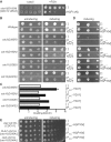
The Hsp40 family member Ydj1 and prefoldin subunit Gim3 are required for septin quality control. (A) As in Figure 1A, but with haploid cells of the indicated genotypes. Strains: BY4741 (wild type); JTY5445 (ydj1∆); MMY0051 (gim3∆). Arrowheads, strong bud neck fluorescence signal. (B) As in Figure 2D, for the strains in A, the hsp104∆ strain JTY4014, the apj1∆ strain MMY0054, the hsp42∆ strain MMY0057, the hsp82∆ strain MMY0058, and the ssb1∆ strain MMY0059. p value, one-tailed unpaired t- test. (C) Cells of the same strains as in A carrying plasmids expressing from the GAL1/10 promoter C-terminally truncated alleles of CDC12 ([PGAL–CDC12∆C]) were resuspended in sterile water and subjected to 10-fold serial dilutions. The residue at position 268 is indicated as wild-type (G; plasmid pMVB160) or substituted to arginine (R; plasmid YCpUG-Cdc12(G268R ∆339-407)). A 5-μl amount of each dilution was spotted on solid synthetic medium containing 10 μg/ml phloxine B to stain dead cells red and dextrose or galactose, as indicated, to repress or induce the GAL1/10 promoter. As indicated, uracil was left out or added to the medium to impose or relieve plasmid selection, respectively. Plates were photographed after 3 d at 30°C.
FIGURE 8:
FIGURE 8:
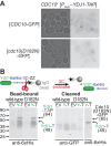
Sequestration of mutant septins by the chaperone Ydj1. (A) Cultures of strain BJ2168 carrying plasmid pLA10K ([CDC10-GFP], wild-type) or YCpK-Cdc10-1-GFP ([cdc10(D182N)-GFP], D182N) and either the empty vector pRS316 (EV) or a plasmid driving TAP-tagged Ydj1 from the GAL1/10-promoter (pPgal-YDJ1, [PGAL–YDJ1-TAP], Y-T) were grown at 30°C in medium lacking uracil and containing 1% raffinose and 1% galactose to induce expression from the GAL1/10 promoter. GFP fluorescence in aliquots of the Ydj1-expressing cells were imaged. (B) The cultures described in A were lysed, and clarified lysates were incubated with IgG-coated beads. After extensive washing, cleaved Ydj1- and Ydj1-bound proteins were eluted from the beads by the addition of GST-tagged 3C protease (3C), which cleaves between the hexahistidine tag (HHHHHH) and IgG-binding domain (ZZ) of Ydj1-TAP, releasing hexahistidine-tagged Ydj1 (Ydj1-T). The beads were boiled in sample buffer (Bead-bound) and, together with the eluate (Cleaved) samples, resolved by SDS–PAGE and transferred to a PVDF membrane. Cdc10(D182N)-GFP was detected using mouse anti-GFP antibodies. Uncleaved Ydj1-TAP and cleaved, hexahistidine-tagged Ydj1 were detected using anti-hexahistidine (anti-6xHis) antibodies (Y1011; UBPBio, Aurora, CO). The leftmost lanes contain a molecular weight ladder (928-60000; Li-Cor Chameleon Duo Pre-stained Protein Ladder). whose contents are labeled on the left with black arrows and sizes. Right, black arrows point to bands migrating with the approximate molecular weight expected for the indicated proteins.
FIGURE 9:
FIGURE 9:
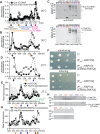
The chaperone Hsp104 sequesters mutant septins in an ATPase-independent manner. (A) Cells of CDC10-SNAP strain JTY4034 (Cdc10-SNAP) or cdc10(D182N)-SNAP strain JTY5168 (Cdc10(D182N)-SNAP) were exposed to 5 μM BG to inactivate the SNAP tags on existing septin–SNAP fusions. After 45 min of subsequent growth at 30°C in the absence of BG, cells were harvested and lysed. Newly synthesized septin-SNAP proteins were fluorescently labeled in the lysate with BG-OG. Lysates were then resolved by SEC. Note that 500 mM NaCl was used to ensure dissociation of septin filaments. Plotted is the BG-OG fluorescence measured for 200-μl portions of each 2-ml fraction. Values shown are normalized to the fraction with the highest signal. Below, filled arrows point to peak fractions classified by color: blue, fluorescence independent of BG-OG addition (Supplemental Figure S2B); magenta, fluorescence requires BG-OG addition, but fractions contain no detectable SNAP-tagged protein (see C); black, predicted molecular weight is consistent with that of septin hetero-octamers; orange, observed in lysates from cdc10(D182N)-SNAP but not CDC10-SNAP cells. (B) As in A, but considering only fractions corresponding to molecular weights >400 kDa. Values were normalized to the fraction with the highest signal in this molecular weight range. (C) Immunoblot analysis of specific fractions from A and B. Fractions are identified by the relevant genotype of the source strain (CDC10, JTY4034; cdc10(D182N)-SNAP, JTY5168) and by predicted molecular weight, using the same color scheme as in A and B. The membrane was exposed to anti–SNAP-Tag antibodies (P9310S; New England Biolabs) and infrared dye–labeled secondary antibodies. After detection (top), the membrane was exposed to anti-Hsp104 antibodies (ADI-SPA-1040-D; Enzo Life Sciences ) and the same secondary antibodies, then scanned again (bottom). The leftmost lane contains a molecular weight ladder (EZ-Run Prestained Protein Marker; Fisher BioReagents, Pittsburgh, PA), whose contents are labeled on the left with black arrows and sizes (in kilodaltons). Black arrows on the right point to bands migrating with the molecular weight expected for the indicated proteins. Gray arrows point to presumed proteolysis products or cross-reacting bands. (D) As in B, but cells were grown at 27°C during the “chase” period. Plotted values represent measurements made from pools of 100 μl of each of two adjacent fractions. Fractions >650 kDa were not measured. (E) As in B and C, but only strain JTY5169 was used, and fluorescence values were not normalized. In the “no BG-OG” sample, no BG-OG was added to the lysate. In the “Hsp104-overexpressed” sample, the cells carried plasmid pRS316-Pgal-HSP104 and were induced to overexpress Hsp104 5.25 h before BG addition. Columns (gray, Hsp104; green, SNAP) indicate levels (in arbitrary units) of Hsp104 or Cdc10(D182N)-SNAP in the indicated fractions from the Hsp104-overexpressing cells as assessed by immunoblotting (Supplemental Figure S2C). The open orange arrow indicates where BG-OG-labeled Cdc10(D182N)-SNAP molecules were detected in D. Red arrow, peak fraction containing BG-OG-labeled Cdc10(D182N)-SNAP only when Hsp104 was overexpressed. (F) As in Figure 6D, with cdc10(D182N) strain CBY06417, plasmid pRS316, pRS316-Pgal-HSP104, or pRS316-PGAL-HSP104(E285Q E687Q), and media lacking histidine. (G) As in E, showing the same data for overexpression of wild-type Hsp104 in strain JTY5169 overlaid with data from overexpression of Hsp104(E285Q E687Q). The yellow arrow indicates a peak fraction of OG fluorescence specific to the Hsp104(E285Q E687Q)-overexpressing sample. (H) As in C, but with fractions from the Hsp104(E285Q E687Q)-overexpressing sample separated on a 4–20% gradient SDS–PAGE gel before immunoblotting and a different molecular weight ladder (Chameleon Duo Pre-stained Protein Ladder, 928-60000; Li-Cor).
FIGURE 10:
FIGURE 10:
Model for chaperone-mediated quality control of higher-order septin assembly. Nascent septin polypeptides emerging from the ribosome encounter a number of cytosolic chaperones during subsequent de novo folding. Those most relevant to quality control are illustrated here. Wild-type septins efficiently adopt quas-inative conformations, thereby burying hydrophobic residues and escaping chaperone-mediated sequestration. Heterodimerization with other septins—the first oligomerization step toward septin filament assembly—occurs concomitant with exit from the chamber of the cytosolic chaperonin CCT (also called TRiC). Mutant septins that inefficiently fold the G heterodimerization interface are slower to achieve a conformation allowing chaperone release. Interactions with the prefoldin complex (PFD), the Hsp40 chaperone Ydj1, and the disaggregase Hsp104 are particularly prolonged, leading to a delay in availability of the mutant septin for hetero-oligomerization. When expressed at endogenous levels, Hsp104 exerts a passive bind-and-release “holdase” function. On overexpression, Hsp104 “unfoldase” activity may be unleashed on the mutant septin, but “holdase” function is sufficient in these conditions to delay mutant septin availability.

Similar articles

Cited by

References

    1. Alpers JB, Paulus H. Allosteric preconditioning: role of allosteric ligands in promoting the maturation of enzymes. Nature. 1971;233:478–480. - PubMed
    1. Amberg D, Burke D, Strathern J. Methods in Yeast Genetics, Cold Spring Harbor, NY: Cold Spring Harbor Laboratory Press; 2005.
    1. Bertin A, McMurray MA, Grob P, Park S-S, Garcia G, 3rd, Patanwala I, Ng H-L, Alber T, Thorner J, Nogales E. Saccharomyces cerevisiae septins: supramolecular organization of heterooligomers and the mechanism of filament assembly. Proc Natl Acad Sci USA. 2008;105:8274–8279. - PMC - PubMed
    1. Betting J, Seufert W. A yeast Ubc9 mutant protein with temperature-sensitive in vivo function is subject to conditional proteolysis by a ubiquitin- and proteasome-dependent pathway. J Biol Chem. 1996;271:25790–25796. - PubMed
    1. Bordallo J, Plemper RK, Finger A, Wolf DH. Der3p/Hrd1p is required for endoplasmic reticulum-associated degradation of misfolded lumenal and integral membrane proteins. Mol Biol Cell. 1998;9:209–222. - PMC - PubMed

Publication types

MeSH terms