. 2015 Apr 15;128(8):1617-26.
doi: 10.1242/jcs.163303. Epub 2015 Feb 11.

Rab11a regulates syntaxin 3 localization and microvillus assembly in enterocytes

Affiliations

Rab11a regulates syntaxin 3 localization and microvillus assembly in enterocytes

Byron C Knowles et al. J Cell Sci. .

Abstract

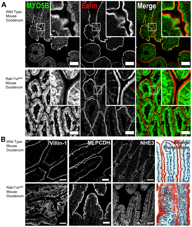
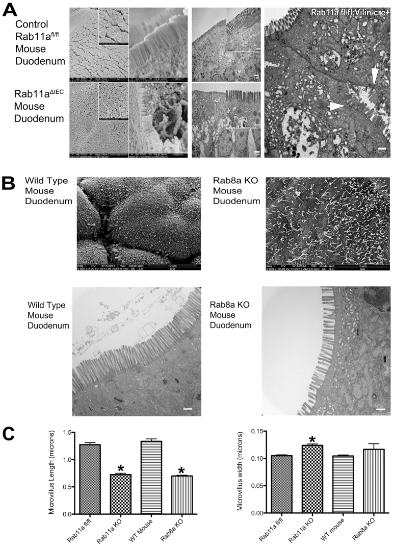
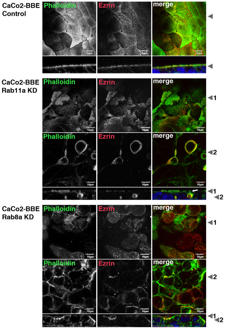
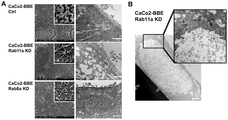
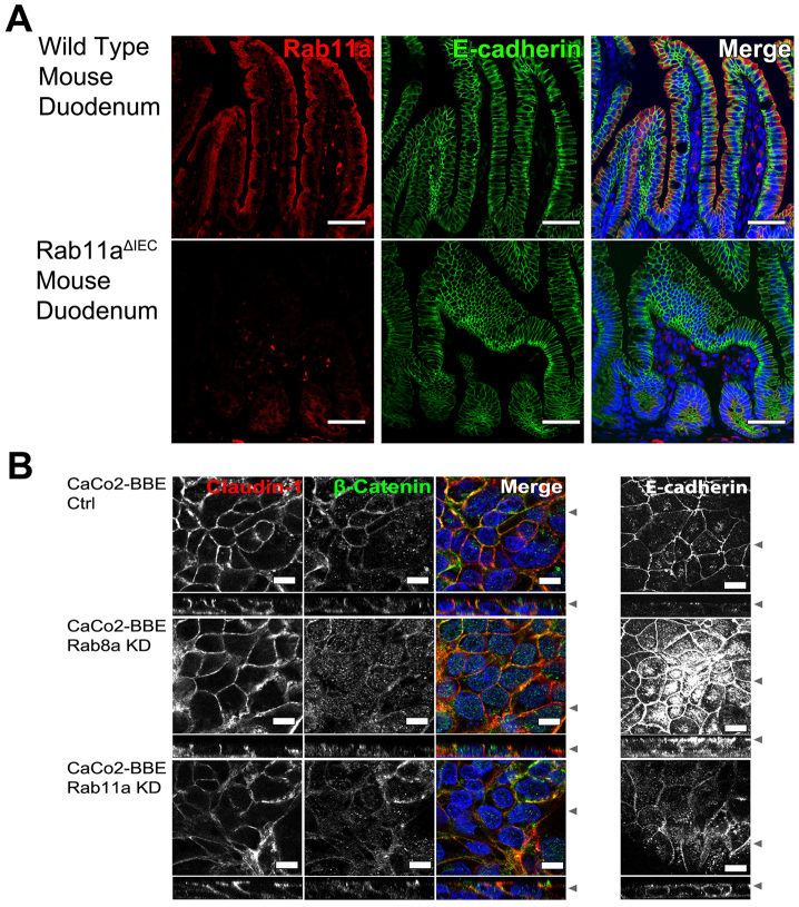
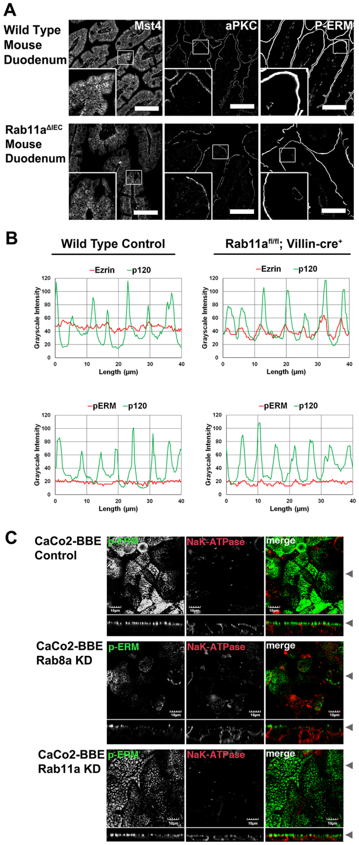
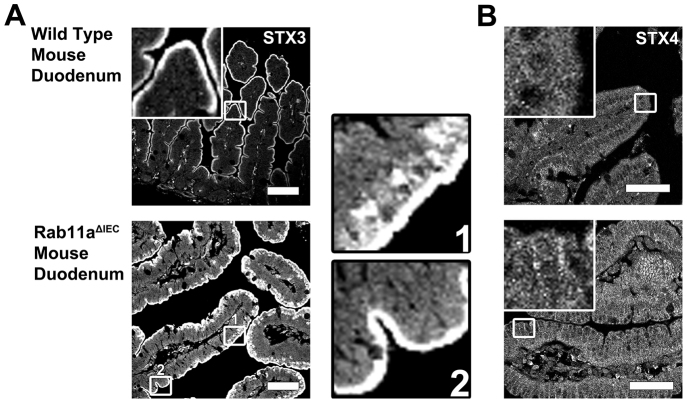
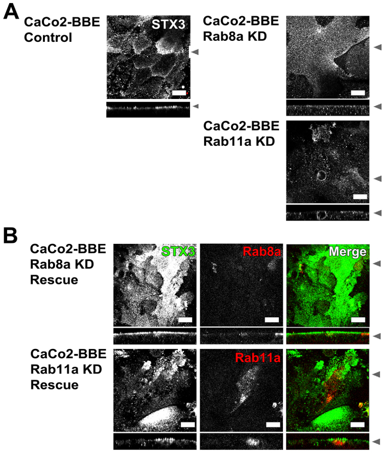
Rab11a is a key component of the apical recycling endosome that aids in the trafficking of proteins to the luminal surface in polarized epithelial cells. Utilizing conditional Rab11a-knockout specific to intestinal epithelial cells, and human colonic epithelial CaCo2-BBE cells with stable Rab11a knockdown, we examined the molecular and pathological impact of Rab11a deficiency on the establishment of apical cell polarity and microvillus morphogenesis. We demonstrate that loss of Rab11a induced alterations in enterocyte polarity, shortened microvillar length and affected the formation of microvilli along the lateral membranes. Rab11a deficiency in enterocytes altered the apical localization of syntaxin 3. These data affirm the role of Rab11a in apical membrane trafficking and the maintenance of apical microvilli in enterocytes.

Keywords: Enterocyte; Intestinal polarity; Microvilli; Rab11; Rab8; Syntaxin 3.


Figures

Fig. 1.
Fig. 1.
Rab11a loss in the enterocytes of mice causes lateral and cytoplasmic redistribution of apical proteins.(A) Control wild-type mouse and Rab11aΔIEC mouse duodenum were dual immunostained fro MYO5B (green) and ezrin (red). White boxes indicate the enlarged regions shown in the insets. In Rab11aΔIEC mouse duodenum, we observed redistribution of MYO5B away from its normal subapical position as well as into the nucleus. Ezrin staining was present on the apical surfaces in both wild-type and Rab11aΔIEC mouse duodenum, but in the Rab11aΔIEC duodenum we also observed staining ezrin along the lateral membranes. (B) Wild-type and Rab11aΔIEC mouse duodenum sections were stained for proteins normally expressed in the enterocyte brush border. Although all four proteins were still associated with the apical membrane, in Rab11aΔIEC mouse duodenum, villin was also present in the lateral membranes and there was an increase in cytosolic NHE3. Both MLPCDH and alkaline phosphatase immunolabeling stained showed increased staining in the sub-apical cytosol, especially in enterocytes at the villus tips in the Rab11aΔIEC mouse duodenum. Scale bars: 30 µm. All results are representative of three separate experiments.
Fig. 2.
Fig. 2.
SEM and TEM of Rab11a- and Rab8a-knockout mice display aberrant intestinal microvilli.(A) SEM and TEM images of control Rab11afl/fl and Rab11aΔIEC mouse duodenum. Both SEM and TEM images of the Rab11aΔIEC mouse duodenum showed short microvilli with normal packing along with large subapical vesicles. The right panel shows enterocytes from Rab11aΔIEC mice demonstrating the presence of microvilli on the lateral membrane (white arrows). Left: Scale bars in SEM: 4 µm, 500 nm for inset, Middle TEM: scale bars are 2 µm and inset 500 nm; left panel scale bar is 500 nm. (B) SEM (first row) and TEM (second row) of wild-type and Rab8a-knockout (KO) mouse duodenum. Rab8a-KO mouse enterocytes displayed sparse brush borders with short microvilli. SEM scale bars: 10 µm; TEM scale bars: 1 µm. (C) Quantification of microvilli dimensions in Rab11aΔIEC and Rab8A-KO mouse enterocytes. Both Rab8a-KO and Rab11aΔIEC mice duodenum display shortened microvilli. Microvilli in Rab11aΔIEC mouse duodenum also showed a significant increase in microvillus width. WT, wild type. Results are mean±s.e.m. *P<0.001. All results are representative of four separate experiments.
Fig. 3.
Fig. 3.
Rab8a-KD and Rab11a-KD in CaCo2-BBE cells recapitulates the phenotype of Rab8a and Rab11a loss in the intestines of mice.phalloidin (green), to visualize F-actin, and ezrin (red). Single channels are shown in grayscale with triple-stained images at left including DAPI nuclear staining (blue). X-Y confocal images are shown above X-Z images. For Rab11a KD and Rab8a KD cells, X-Y images are shown for both an apical region (arrowhead 1 in X-Z) and a medial region (arrowhead 2 in X-Z). Arrowheads at the right in X-Y images indicate the position of the corresponding X-Z image. In control cells, ezrin staining was confined to the apical membrane. F-actin staining was concentrated at the apical membrane with fainter staining observed along the lateral membrane. Rab11a-KD cells displayed reduced apical ezrin and phalloidin staining, but also showed bright phalloidin and ezrin staining in regions along lateral membranes. Note the region indicated by a white arrow, where lateral staining, which is positive only for F-actin, surmounts an area of dual staining for ezrin and F-actin along the lateral membranes. These images are consistent with the observation of lateral microvilli. Rab8a KD cells showed a reduction in apical F-actin and ezrin staining, but did not show lateral ezrin staining. All results are representative of three separate experiments. Scale bars: 10 µm.
Fig. 4.
Fig. 4.
SEM and TEM examination of Rab11a KD and Rab8a KD cells demonstrates deficits in apical microvilli.-KD and Rab8a-KD cells were examined by SEM and TEM. (A) Rab8a KD cells demonstrated more immature and sparser microvilli than Rab11a KD cells. Scale bars are as indicated. (B) TEM of enlarged lateral spaces containing microvilli in CaCo2-BBE Rab11a-KD cell. Scale bars: 2 µm (main panel); 500 nm (inset). All results are representative of four separate experiments.
Fig. 5.
Fig. 5.
Rab11a loss does not cause loss of basolateral integrity in enterocytes.(A) Sections of wild-type and Rab11aΔIEC mouse duodenum were stained for Rab11a (red) and E-cadherin (green), with the merged image at the right including DAPI nuclear stain (blue). In Rab11aΔIEC mouse duodenum, E-cadherin was maintained in the basolateral compartment of enterocytes, but the cells did appear to lose some contact inhibition. Scale bars: 20 µm. (B) Left panels: control CaCo2-BBE, Rab8a KD and Rab11a KD cells were stained for claudin-1 (red) and β-catenin (green) with the merged image shown at right including DAPI nuclear stain (blue). In control cells, claudin-1 and β-catenin were distributed along the basolateral surface. In Rab8a-KD cells, claudin-1 was maintained at its basolateral position, but β-catenin was shifted to a cytoplasmic localization. In Rab11a-KD cells, claudin-1 and β-catenin were distributed along the basolateral surface. Right panels: cells were stained for E-cadherin. In control cells, E-cadherin was positioned in a junctional localization. In Rab8a KD cells, E-cadherin was accumulated in the cytosol, but was still present on the lateral membranes. In Rab11a-KD cells, E-cadherin was redistributed to both the apical and basolateral surfaces. Arrowheads at the right in X-Y images indicate the position of the corresponding X-Z image. Scale bars: 10 µm. All results are representative of three separate experiments.
Fig. 6.
Fig. 6.
Rab11aΔIEC mouse duodenum samples display apical localization of aPKC and phosphorylated ERM proteins.(A) Sections of wild-type and Rab11aΔIEC duodenum were stained for Mst4, aPKC and P-ERM. In wild-type mouse duodenum, Mst4 was localized to the cytoplasm and showed a distinct sub-apical concentration, but in Rab11aΔIEC duodenum samples the sub-apical population was lost. aPKC and P-ERM proteins were localized to the apical surface in both wild-type and Rab11aΔIEC duodenum samples. Scale bars: 50 µm. (B) To analyze the presence of ezrin and P-ERM along the lateral membranes of wild-type and Rab11aΔIEC enterocytes, staining for both were compared with lateral membrane p120 catenin staining using linear intensity determinations. Representative tracings demonstrate that although ezrin was present along the lateral membranes of Rab11aΔIEC enterocytes, little P-ERM immunoreactivity could be detected. (C) In CaCo2-BBE cells, immunostaining for the lateral membrane marker Na+/K+-ATPase (red) was compared with P-ERM staining (green). Individual channels are shown in grayscale with the merged dual-label image shown on the right. In control CaCo2-BBE cells, phosphorylated ERM proteins were distributed tightly along the apical surface. In Rab8a-KD cells and Rab11a-KD cells, phosphorylated ERM proteins were maintained at the apical membrane. Arrowheads at the right in X-Y images indicate the position of the corresponding X-Z image. Scale bars: 10 µm. All results are representative of three separate experiments.
Fig. 7.
Fig. 7.
Loss of Rab11a alters the apical localization of STX3 in enterocytes.(A) Sections of wild-type and Rab11aΔIEC duodenum were stained for STX3. In wild-type mouse duodenum, STX3 was localized to the apical surface. In Rab11aΔIEC mice duodenum, STX3 was reorganized away from the apical surface in punctate vesicles below the apical surface. Insets indicated by numbers 1 (top) and 2 (bottom) display redistribution of STX3 in enterocytes at the villus tips, whereas enterocytes towards the bases of villi showed a normal apical distribution. (B) In both wild-type and Rab11aΔIEC mice duodenum STX4 was localized to intracellular vesicles along the lateral membrane. Scale bars: 50 µm. All results are representative of three separate experiments.
Fig. 8.
Fig. 8.
Both MYO5B and Rab11a are required for proper localization of STX3.(A) CaCo2-BBE cell lines were stained for STX3. In control cells, STX3 was localized to the apical surface. In Rab8a-KD cells, STX3 accumulated in vesicles below the apical surface. In Rab11a-KD cells, STX3 was redistributed away from the apical surface into the cytoplasm and to lateral membranes. Scale bars: 10 µm. (B) STX3 immunostaining (green) was evaluated in Rab8a KD cells with mCherry–Rab8a rescue (red) or Rab11a KD cells with mCherry–Rab11a rescue (red). Rab8a re-expression in Rab8a-KD re-established discrete apical localization of STX3. Rab11a re-expression in Rab11a-KD cells, re-established the normal apical localization of STX3. Arrowheads at the right in X-Y images indicate the position of the corresponding X-Z image. Scale bars: 10 µm. All results are representative of three separate experiments.

Similar articles

Cited by

References

    1. Baetz NW, Goldenring JR. Rab11-family interacting proteins define spatially and temporally distinct regions within the dynamic Rab11a-dependent recycling system. Mol. Biol. Cell. 2013;24:643–658. doi: 10.1091/mbc.E12--09--0659. - DOI - PMC - PubMed
    1. Barnard JA, Beauchamp RD, Coffey RJ, Moses HL. Regulation of intestinal epithelial cell growth by transforming growth factor type beta. Proc. Natl. Acad. Sci. USA. 1989;86:1578–1582. doi: 10.1073/pnas.86.5.1578. - DOI - PMC - PubMed
    1. Bryant DM, Datta A, Rodríguez-Fraticelli AE, Peränen J, Martín-Belmonte F, Mostov KE. A molecular network for de novo generation of the apical surface and lumen. Nat. Cell Biol. 2010;12:1035–1045. doi: 10.1038/ncb2106. - DOI - PMC - PubMed
    1. Calhoun BC, Lapierre LA, Chew CS, Goldenring JR. Rab11a redistributes to apical secretory canaliculus during stimulation of gastric parietal cells. Am. J. Physiol. 1998;275:C163–C170. - PubMed
    1. Dhekne HS, Hsiao NH, Roelofs P, Kumari M, Slim CL, Rings EH, van Ijzendoorn SC. Myosin Vb and Rab11a regulate phosphorylation of ezrin in enterocytes. J. Cell Sci. 2014;127:1007–1017. doi: 10.1242/jcs.137273. - DOI - PubMed

Publication types

LinkOut - more resources