

Flow cytometric evaluation of disseminated intravascular coagulation in a canine endotoxemia model
- PMID: 25673909
- PMCID: PMC4283234
Flow cytometric evaluation of disseminated intravascular coagulation in a canine endotoxemia model
Abstract
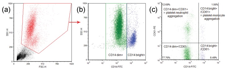
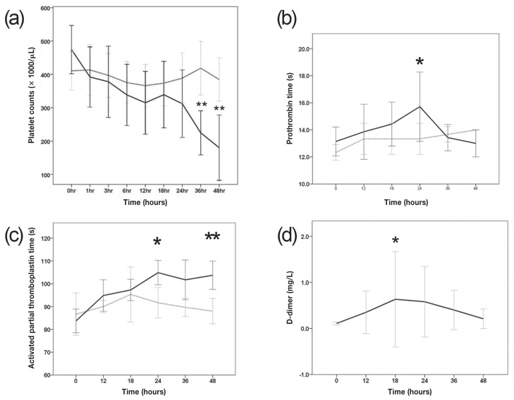
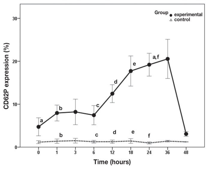
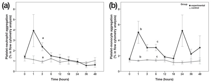
Sepsis is associated with substantial morbidity and mortality in dogs. Alterations in hemostasis by systemic inflammation play an important role in the pathophysiology of sepsis. To evaluate the functional hemostatic changes in sepsis, we evaluated coagulation profiles and flow cytometric measurement of P-selectin (CD62P) expression on platelets, as well as platelet-leukocyte aggregation from a lipopolysaccharide (LPS)-induced endotoxemia model in dogs (n = 7). A sublethal dose of LPS [1 mg/kg body weight (BW)] induced thrombocytopenia and increased activated partial thromboplastin time (aPTT), prothrombin time (PT), and D-dimer concentrations. Flow cytometry analysis showed a significant increase in P-selectin expression on platelets between 1 and 24 h of a total 48 h of the experiment. In addition, platelet-leukocyte aggregation was significantly increased in the early stage of endotoxemia (at 1 and < 6 h for platelet-monocyte aggregation and at 3 h for platelet-neutrophil aggregation). Our results suggest that CD62P expression on platelets and platelet-leukocyte aggregation, as measured by flow cytometry, can be useful biomarkers of disseminated intravascular coagulation (DIC) in canine sepsis. These functional changes contribute to our understanding of the pathophysiology of hemostasis in endotoxemia.
Chez les chiens la septicémie est associée à une morbidité et une mortalité élevée. Les modifications de l’hémostase par une inflammation systémique jouent un rôle important dans la pathophysiologie de la septicémie. Afin d’évaluer les changements hémostatiques fonctionnels lors de septicémie, une évaluation fut faite des profils de coagulation et des mesures par cytométrie en flux de l’expression de P-sélectine (CD62) sur les plaquettes, ainsi que de l’agrégation plaquettes-leucocytes dans un modèle d’endotoxémie induite par le lipopolysaccharide (LPS) chez des chiens (n = 7). Une dose sub-léthale de LPS [1 mg/kg de poids corporel] induisit une thrombocytopénie et augmenta le temps de thromboplastine partielle activée (aPTT), le temps de prothrombine (PT), et les concentrations de dimère-D. L’analyse par cytométrie en flux a montré une augmentation significative de l’expression de P-sélectine sur les plaquettes entre 1 et 24 h du total de 48 h que dura l’expérience. De plus, l’agrégation plaquettes-leucocytes était augmentée de manière significative dans les stages initiaux de l’endotoxémie (à 1 et < 6 h pour l’agrégation plaquettes-monocytes et 3 h pour l’agrégation plaquettes-neutrophiles). Nos résultats suggèrent que l’expression de CD62P sur les plaquettes et l’agrégation plaquettes-leucocytes, telle que mesurée par cytométrie en flux, peuvent être des biomarqueurs utiles de la coagulation intravasculaire disséminée (DIC) lors de septicémie canine. Ces changements fonctionnels contribuent à notre compréhension de la pathophysiologie de l’hémostase lors d’endotoxémie.(Traduit par Docteur Serge Messier).
Figures
References
-
- Levy MM, Fink MP, Marshall JC, Abraham E, Angus D, Cook D, et al. 2001 SCCM/ESICM/ACCP/ATS/SIS International Sepsis Definitions Conference. Intensive Care Med. 2003 Apr;29:530–538. - PubMed
-
- Bentley AM, Otto CM, Shofer FS. Comparison of dogs with septic peritonitis: 1988–1993 versus 1999–2003. J Vet Emerg Crit Care (San Antonio) 2007;17:391–398.
-
- Yu D-H, Nho D-H, Song R-H, et al. High-mobility group box 1 as a surrogate prognostic marker in dogs with systemic inflammatory response syndrome. J Vet Emerg Crit Care (San Antonio) 2010;20:298–302. - PubMed
-
- Rau S, Kohn B, Richter C, et al. Plasma interleukin-6 response is predictive for severity and mortality in canine systemic inflammatory response syndrome and sepsis. Vet Clin Pathol. 2007 Sep;36:253–260. - PubMed
-
- Luschini MA, Fletcher DJ, Schoeffler GL. Retrospective Study: Incidence of ionized hypocalcemia in septic dogs and its association with morbidity and mortality: 58 cases (2006–2007) J Vet Emerg Crit Care. 2010;20:406–412. - PubMed
Publication types
MeSH terms
Substances
LinkOut - more resources
Full Text Sources