Randomized Controlled Trial
. 2015 Feb 2:10:387-403.
doi: 10.2147/CIA.S69752. eCollection 2015.

Mécano-Stimulation™ of the skin improves sagging score and induces beneficial functional modification of the fibroblasts: clinical, biological, and histological evaluations

Affiliations
Randomized Controlled Trial

Mécano-Stimulation™ of the skin improves sagging score and induces beneficial functional modification of the fibroblasts: clinical, biological, and histological evaluations

Philippe Humbert et al. Clin Interv Aging. .

Abstract

Background: Loss of mechanical tension appears to be the major factor underlying decreased collagen synthesis in aged skin. Numerous in vitro studies have shown the impact of mechanical forces on fibroblasts through mechanotransduction, which consists of the conversion of mechanical signals to biochemical responses. Such responses are characterized by the modulation of gene expression coding not only for extracellular matrix components (collagens, elastin, etc.) but also for degradation enzymes (matrix metalloproteinases [MMPs]) and their inhibitors (tissue inhibitors of metalloproteinases [TIMPs]). A new device providing a mechanical stimulation of the cutaneous and subcutaneous tissue has been used in a simple, blinded, controlled, and randomized study.

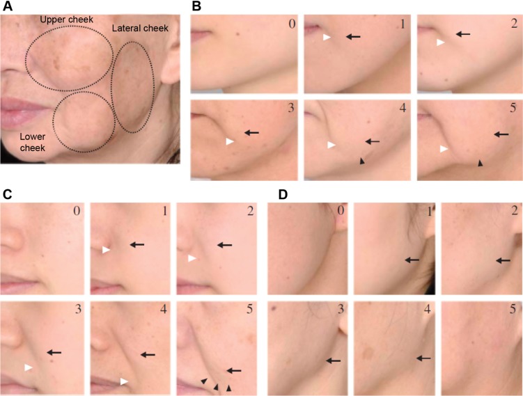
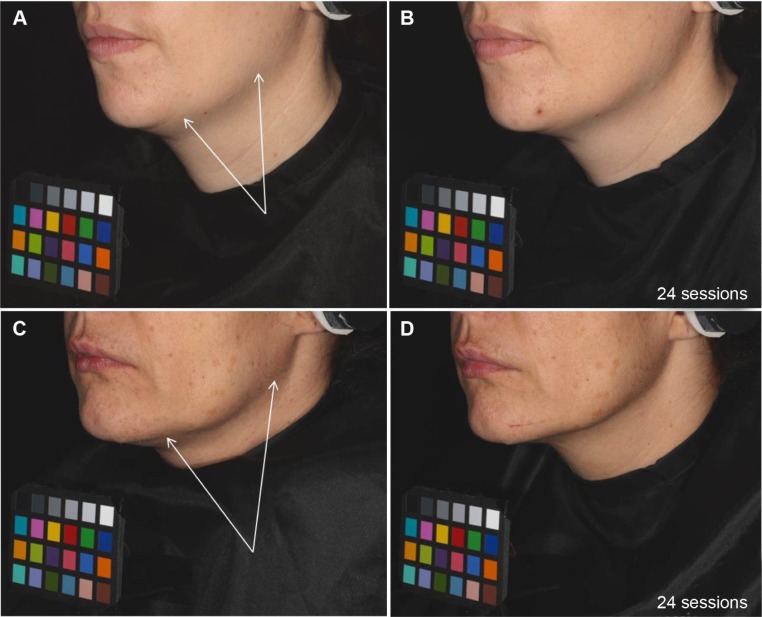
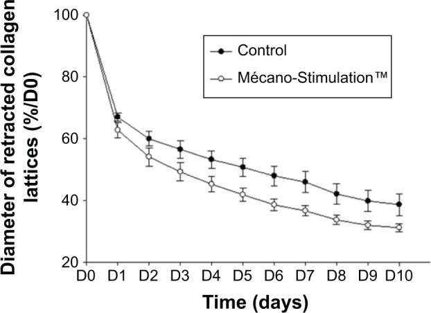
Materials and methods: Thirty subjects (aged between 35 years and 50 years), with clinical signs of skin sagging, were randomly assigned to have a treatment on hemiface. After a total of 24 sessions with Mécano-Stimulation™, biopsies were performed on the treated side and control area for in vitro analysis (dosage of hyaluronic acid, elastin, type I collagen, MMP9; equivalent dermis retraction; GlaSbox(®); n=10) and electron microscopy (n=10). Furthermore, before and after the treatment, clinical evaluations and self-assessment questionnaire were done.

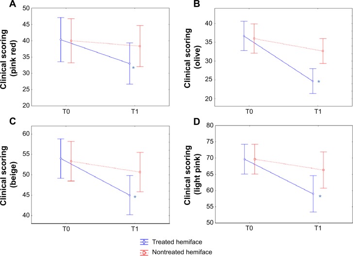
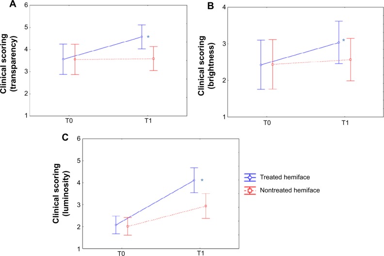
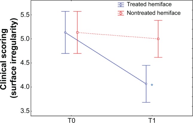
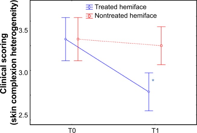
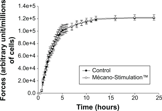
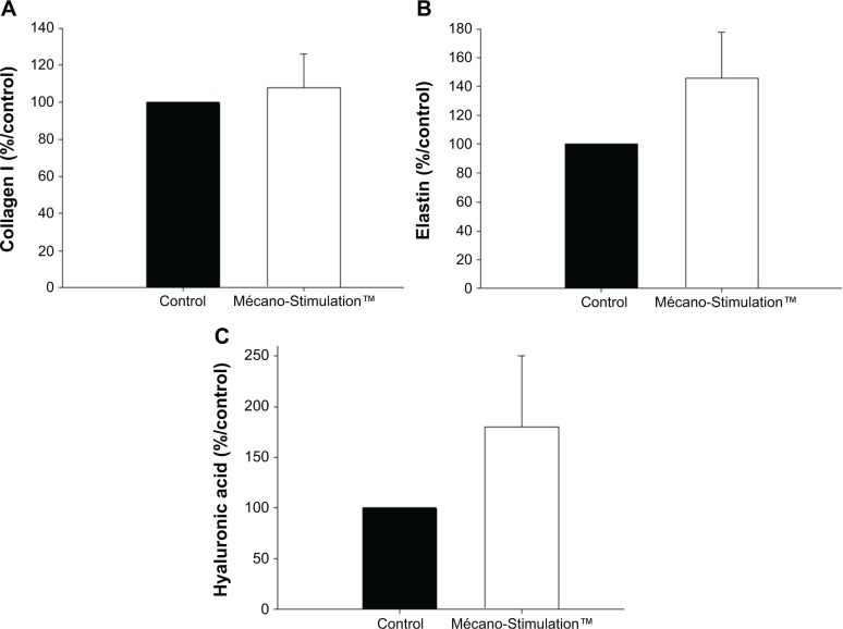
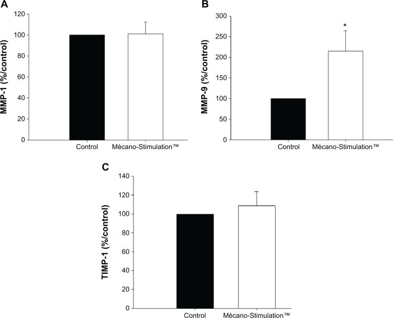
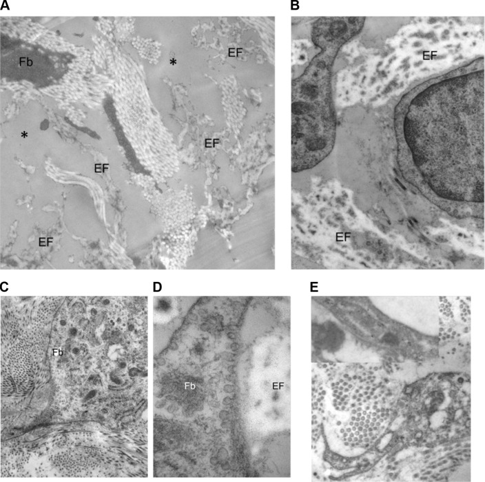
Results: In vitro analysis showed increases in hyaluronic acid, elastin, type I collagen, and MMP9 content along with an improvement of the migratory capacity of the fibroblasts on the treated side. Electron microscopy evaluations showed a clear dermal remodeling in relation with the activation of fibroblast activity. A significant improvement of different clinical signs associated with skin aging and the satisfaction of the subjects were observed, correlated with an improvement of the sagging cheek.

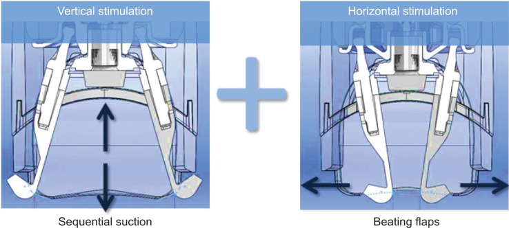
Conclusion: Mécano-Stimulation is a noninvasive and safe technique delivered by flaps microbeats at various frequencies, which can significantly improve the skin trophicity. Results observed with objective measurements, ie, in vitro assessments and electron microscopy, confirm the firming and restructuring effect clinically observed.

Keywords: fibroblast synthesis; mechanical stimulation; skin rejuvenation; skin sagging.


Figures

Figure 1
Figure 1
Ezure scoring scale. Notes: Skin sagging is evaluated through the Ezure scoring scale: facial cutaneous sagging is graded from 0 to 5 on three areas of the hemiface (A), upper cheek (B), lower cheek (C), and lateral cheek (D). Arrows and arrowheads point out specific areas of interest defining skin sagging stages. Volunteers selected in the study were of grade 1–3.
Figure 2
Figure 2
Double stimulation. Notes: The Mécano-Stimulation™ technique consists of two motorized flaps located inside a treatment chamber and dedicated to the treatment of the face or small areas. The treatment forces combine sequential aspiration and beating flaps creating, respectively, vertical and horizontal stimulation. Settings allow obtaining different intensity and rhythmicity of the aspiration or beating frequency of the flaps. Lift heads used in this study were “motorized”: heads contain a small engine allowing the flaps to beat in a regular and permanent way, synchronized specifically with the sequential aspiration.
Figure 3
Figure 3
CLBT™ scoring before (T0) and after (T1) the 24 treatment sessions: results for the color components. Notes: The visual analysis scale (CLBT scoring12) follows seven descriptors that evaluate between 10% and 100% of four different colors: pink red (A), olive (B), beige (C), and light pink (D). All these color parameters presented a significant improvement (decrease) in terms of time/treatment evolution, in favor of the treated (blue) versus nontreated (red) side. *P<0.05. Abbreviations: CLBT, Color, Luminosity, Brightness and Transparency Scoring; T, time.
Figure 4
Figure 4
CLBT™ scoring before (T0) and after (T1) the 24 treatment sessions: results for transparency, brightness, and luminosity. Notes: The visual analysis scale (CLBT scoring12) evaluates three parameters of skin transparency (A), brightness (B), and luminosity (C) on a 0–10 scale; a significant improvement (increase) in terms of time/treatment evolution is pictured in all cases, in favor of the treated (blue) versus nontreated (red) hemiface. *P<0.05. Abbreviations: CLBT, Color, Luminosity, Brightness and Transparency Scoring; T, time.
Figure 5
Figure 5
Clinical scoring of skin radiance: relief surface irregularity. Notes: An analogical scale (from 0 to 9) was used to score the radiance through relief parameters: among those, surface irregularity was evaluated from small (0) to large (9) before (T0) and after (T1) the treatment. The figure represents a significant improvement (decrease) in terms of time/treatment evolution observed; surface irregularity score was improved by 20.7% on treated side (blue) against 2.5% on nontreated side (red). *P<0.05. Abbreviation: T, time.
Figure 6
Figure 6
Clinical scoring of skin radiance: color skin complexion heterogeneity. Notes: The scoring of the radiance was also assessed through color parameters: skin complexion heterogeneity score was evaluated with an analogical scale (from 0= homogeneous to 9= heterogeneous) and is presented before (T0) and after (T1) the 24 sessions of treatment. At T1, the score was significantly improved (decrease) on the treated side (blue) compared to the nontreated hemiface (red); a significant improvement in terms of time/treatment evolution was observed. Skin complexion heterogeneity score was improved by 13.8% on the treated side while a worsening was noticed on the nontreated side (+1.2%). *P<0.05. Abbreviation: T, time.
Figure 7
Figure 7
Skin sagging evolution during the study: examples on two volunteers treated on the left side of the face before and after 24 sessions of treatment. Notes: Standardized photographs illustrated an improvement of the face oval: Two examples are pictured: before treatment (A, C) where skin sagging is indicated with arrows on lateral and lower cheek; after 24 sessions of treatment, and (B, D) skin slackening is reduced.
Figure 8
Figure 8
Effect of Mécano-Stimulation™ on fibroblast retraction according to time. Notes: Fibroblasts capacity to retract free-floating collagen lattices reflects their migration, and is presented to time. The retraction of collagen lattices obtained with fibroblasts exposed to Mécano-Stimulation™ was significantly increased (decreasing of the diameter of retracted collagen lattices) compared to nonstimulated fibroblasts (P<0.001).
Figure 9
Figure 9
Quantification of contractile forces developed by fibroblasts after the action of Mécano-Stimulation™ using GlaSbox® device. Notes: The GlaSbox® device contains eight rectangular culture wells in which lattices have been developed; two opposite silicon beams with strain gauges allow force measurements to be obtained. The results are expressed as contractile forces (arbitrary unit) developed by fibroblasts subjected or not subjected to Mécano-Stimulation™ according to the time. No significant difference of the development of contractile forces was observed between Mécano-Stimulation group and control group.
Figure 10
Figure 10
Collagen I, elastin, and hyaluronic acid synthesis of fibroblasts with and without the action of Mécano-Stimulation™. Notes: The quantification of collagen I and hyaluronic acid was evaluated using an enzyme-linked immunosorbent assay kit. The concentration in the samples was determined by comparing the optical density of the samples to the standard curve. The total soluble elastin was assayed by a colorimetric method (Fastin Elastin assay). Results are expressed as % respective control. At T1, nonsignificant increase in the synthesis of collagen I (A) was observed between the treated and untreated hemiface (107.8%±69.8% and 100.0%±0.0%/control). At T1, an increase in the synthesis of elastin (B) and hyaluronic acid (C) was observed between the treated and untreated hemiface (P=0.175 and P=0.268, respectively). Abbreviation: T, time.
Figure 11
Figure 11
MMP-1, MMP-9, and TIMP-1 synthesis of fibroblasts with and without the action of Mécano-Stimulation™. Notes: The quantification of MMP-1, MMP-9, and TIMP-1 was obtained using an enzyme-linked immunosorbent assay kit. Results are expressed as % respective control. No significant change was observed in the synthesis of MMP-1 (A) and TIMP-1 (C). At T1, a significant increase in the synthesis of MMP-9 (B) was observed between the treated and untreated hemiface (P=0.033). *P<0.05 versus control. Abbreviations: MMP, matrix metalloproteinase; TIMP, tissue inhibitor of metalloproteinases; T, time.
Figure 12
Figure 12
Examples of ultrastructure observed when comparing the treated versus untreated side. Notes: Examples of ultrastructure of the papillary dermis observed when comparing the treated versus untreated side. Untreated side (A): Presence of an amorphous substance (*) in the spaces between fragmented EFs. Treated side (B, C, D, and E). (B) Restructured EFs in the vicinity of Fb with clear cytoplasm and developed mitochondria. Fb express well-developed rough endoplasmic network (C) and plasma membrane–attached small secretory vesicles often facing the EFs (D). (E) Immunogold labeling of α-smooth muscle actin in the Fb of the papillary dermis. Abbreviations: EFs, elastic fibers; Fb, fibroblasts.

References

    1. Courderot-Masuyer C, Robin S, Tauzin H, Humbert P. Evaluation of the behaviour of wrinkles fibroblasts and normal aged fibroblasts in the presence of poly-l-lactic acid. J Cosmet Dermatol Sci Appl. 2012;2:20–27.
    1. Jacquet L. Le massage plastique à double action dans le traitement des dermatoses [The double action plastic massage in the treatment of dermatosis] Paris Médical: la semaine du clinicien. 1911;01:355–358. French.
    1. Eastwood M, MacGrouther DA, Brown RA. Fibroblasts responses to mechanical forces. Proc Inst Mech Eng. 1998;212:85–92. - PubMed
    1. Chiquet M. Regulation of extracellular matrix gene expression by mechanical stress. Matrix Biol. 1999;18:417–426. - PubMed
    1. Harris AK, Stopak D, Wild P. Fibroblast traction as a mechanism for collagen morphogenesis. Nature. 1981;290:249–251. - PubMed

Publication types