Review
. 2015 Jan 26:5:682.
doi: 10.3389/fimmu.2014.00682. eCollection 2014.

Unraveling the Interaction between FcRn and Albumin: Opportunities for Design of Albumin-Based Therapeutics

Affiliations
Review

Unraveling the Interaction between FcRn and Albumin: Opportunities for Design of Albumin-Based Therapeutics

Kine Marita Knudsen Sand et al. Front Immunol. .

Abstract

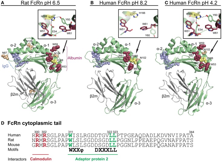
The neonatal Fc receptor (FcRn) was first found to be responsible for transporting antibodies of the immunoglobulin G (IgG) class from the mother to the fetus or neonate as well as for protecting IgG from intracellular catabolism. However, it has now become apparent that the same receptor also binds albumin and plays a fundamental role in homeostatic regulation of both IgG and albumin, as FcRn is expressed in many different cell types and organs at diverse body sites. Thus, to gain a complete understanding of the biological function of each ligand, and also their distribution in the body, an in-depth characterization of how FcRn binds and regulates the transport of both ligands is necessary. Importantly, such knowledge is also relevant when developing new drugs, as IgG and albumin are increasingly utilized in therapy. This review discusses our current structural and biological understanding of the relationship between FcRn and its ligands, with a particular focus on albumin and design of albumin-based therapeutics.

Keywords: FcRn; IgG; albumin; albumin-based therapeutics; half-life; recycling; transcytosis.


Figures

Figure 1
Figure 1
Crystallographic illustrations of rat and human FcRn. Crystal structures of truncated (A) rat FcRn solved at pH 6.5, (B) human FcRn solved at pH 8.2, and (C) pH 4.2. The FcRn HC is shown in green and the β2m subunit in gray (A–C). The three domains of FcRn are denoted as α1, α2, and α3. The key amino acid residues involved in binding to IgG are shown as blue spheres (L112, E115, E116, W131, P132, and E133, human numbering), while the residues central for albumin binding (W51, W53, W59, and W61) are shown as red spheres. The loop containing the tryptophans is pH dependently regulated by H166 (H168 in rat) within the α2-domain (yellow spheres, human numbering). The close-ups show how H166 (H168 in rat) stabilizes the loop of amino acid residues corresponding to residues 51–61 of the α1-domain by forming charged interactions with E54 and Y60 at acidic pH. These interactions are not seen in the crystal structure solved at basic pH, which results in an unstructured loop (B). The four putative N-glycosylation sites of rat FcRn are shown in orange spheres (N87, 104, 128, and 225), while only one N-glycosylation site is found in human FcRn (N102). (D) An amino acid sequence alignment of the cytoplasmic tail of FcRn from human, rat, and mouse. The tryptophan and the di-leucine based sorting motifs that interact with the adaptor protein 2 are highlighted in green. Amino acid residues required for calmodulin binding are marked in red (human numbering). The figures were made using PyMol, with the following PDB files; Rat FcRn pH 6.5: 3FRU (59), human FcRn pH 8.2 1EXU (60), human FcRn pH 4.2: 3MIB (61).
Figure 2
Figure 2
The crystal structure of human albumin. The illustration shows the crystal structure of human albumin solved in the presence of saturating amounts of palmitic acid. The α-helical structures of the three domains (DI, DII, and DIII) are divided into subdomains (A and B) as indicated. DI (pink) contains the fatty acid binding site 1, the free cysteine (C34), and drug binding site 3. Fatty acid site 2 is located at the interface between DI and DII. The metal binding site is located between subdomain DIA and DIIA. DII (orange) contains the drug binding site 1 (Sudlow’s site 1) as well as fatty acid sites 6 and 7. DIII (blue) contains fatty acid binding sites 3 and 4, the drug binding site 2 (Sudlow’s site 2) in DIIIA, and the fatty acid biding site 5 in DIIIB. Examples of the binding sites for endogenous and exogenous ligands for which crystal structures have been solved are listed in green and red, respectively, as reviewed in Ref. (86). The figure was designed using PyMol and the crystal structure data of human albumin solved in the presence of palmitic acid with the PDB file 1E7H (87). CMPF, carboxy-4-methyl-5-propyl-2-furanpropionic acid; NO, nitric oxide.
Figure 3
Figure 3
The co-crystal structure of human FcRn bound to IgG–Fc and albumin. The illustration shows the ternary complex of human FcRn in complex with IgG Fc and wild-type human albumin. The three domains of the FcRn HC (α1, α2, and α3) are shown in green and the β2m subunit in gray. The IgG–Fc is shown in blue while the three domains of albumin are shown in pink (DI), orange (DII), and light blue (DIII). H166 that stabilizes the structure of the loop containing W51, W53, W59, and W61 is shown in yellow. The FcRn residues W53 and W59 are shown as red spheres. (A) FcRn–W59 makes contacts with a hydrophobic pocket in DIIIA, which is composed of T467, T422, V426, L460, L463, and H464. (B) FcRn–W53 makes hydrophobic stacking with three phenylalanines in DIIIB (F507, F509, and F551). (C) A close-up of the structural areas of DI with N111 and N109 of albumin DI interacting with the FcRn residues S58 and K63, respectively. (D) A close-up of the structural interface showing the intramolecular hydrogen bond between albumin R81 and D89, and the interaction between FcRn–T153 and R81. The figures were made using PyMol and the crystal structure data of human FcRn in complex with IgG–Fc and albumin (4N0U) (69).
Figure 4
Figure 4
FcRn-mediated transport pathways. (A) A schematic illustration of the model of FcRn-mediated recycling pathway of its two ligands in an endothelial cell lining the vascular space. (1) IgG and albumin are taken up from the blood by pinocytosis in Rab5 positive early endosomes. (2) FcRn, predominantly localized to acidified endosomes, binds the ligands in Rab4 and Rab11 positive recycling compartments. (3) The ternary complex is recycled to the cell surface as Rab11-positive tubules, which results in exocytosis of the ligands. (4) The neutral pH of the bloodstream leads to release of the ligands. (5) Proteins that do not bind to the receptor will be sorted to late endosomes and further to lysosomes for degradation. (B) An illustration of a polarized epithelial cell layer and the model of FcRn-mediated bidirectional transport. (1) The acidic pH present at certain mucosal sites (apical side) may result in binding of the ligands to FcRn at the cell surface in addition to within recycling endosomes. (2) The transcytotic pathway may be regulated by calmodulin that binds to the cytoplasmic tail of FcRn, and (3) depends on the actin motor myosin Vb and Rab25. (4) Endosomes fuse with the basolateral side of the cells, which lead to release of the ligands upon exposure to neutral pH. (5) FcRn may also transcytose IgG-containing immune complexes across the polarized cell layer. (C) The illustration shows a DC that expresses both FcRn and classical Fcγ receptors. (1) Cross-binding of an IgG-containing immune complex to surface-expressed FcγRs leads to internalization into early endosomes. (2) The immune complexes engage FcRn within acidified endosomes. (3) FcRn directs the IgG-containing immune complexes to loading compartments for processing followed by loading of antigenic peptides onto MHC I (in terms of cross-presentation) and MHC II. (4) MHC I and II traffic to the plasma membrane for presentation of peptides to T-cells. (5) Monomeric ligands may also be recycled by DCs.
Figure 5
Figure 5
Transport of IgG and albumin in the kidneys. A schematic cartoon showing the nephron of the kidney and transport pathways. Blood enters the kidneys through the afferent arteriole from the renal artery, proceeds through the capillaries of the glomerulus where filtration occurs, and exits through the efferent arteriole. (A) The filtration barrier of the glomerulus. (1) The first barrier is the fenestrations between endothelial cells of the glomerular capillaries. (2) The second barrier is the basement membrane, a non-cellular layer consisting of extracellular matrix molecules, which make up charged pores. The podocytes are specialized epithelial cells that encapsulate the capillaries and the basement membrane, and form the outermost layer of epithelial cells facing the glomerular filtrate. (3) The foot processes of the podocytes have slits between them (slit diaphragms) that form the third layer of the filtration barrier. (4) As the pore size of the barrier is between 60–70 kDa, close to the size of albumin (66.5 kDa), some albumin passes the filter. (5) Podocytes express FcRn, and may transcytose IgG and albumin to the glomerular filtrate to prevent clogging of the filter. (B) The glomerular filtrate enters the proximal tubuli where proximal tubular epithelial cells lining the lumen of the tubuli are involved in reabsorption of albumin and IgG, and thus prevent loss into the urine. (1) Proximal tubular epithelial cells also express the cubilin/megalin receptor complex that binds albumin. (2) In acidified endosomes, FcRn binds the ligands, and facilitate transcytosis. (3) The ligands are delivered to the interstitial space of the kidneys followed by drainage to lymphatic vessels and re-entry to the blood circulation.

References

    1. Waldmann TA, Strober W. Metabolism of immunoglobulins. Prog Allergy (1969) 13:1–110. - PubMed
    1. Peters T. Serum albumin. Adv Protein Chem (1985) 37:161–245 10.1016/S0065-3233(08)60065-0 - DOI - PubMed
    1. Spiegelberg HL, Fishkin BG. The catabolism of human G immunoglobulins of different heavy chain subclasses. 3. The catabolism of heavy chain disease proteins and of Fc fragments of myeloma proteins. Clin Exp Immunol (1972) 10(4):599–607. - PMC - PubMed
    1. Peters T. All about albumin: Biochemistry, Genetics and Medical Applications. San Diego CA: Academic Press; (1996).
    1. Kragh-Hansen U, Chuang VT, Otagiri M. Practical aspects of the ligand-binding and enzymatic properties of human serum albumin. Biol Pharm Bull (2002) 25(6):695–704. 10.1248/bpb.25.695 - DOI - PubMed