

Thoracic extraosseous epidural cavernous hemangioma
- PMID: 25674348
- PMCID: PMC4323510
- DOI: 10.3340/jkns.2015.57.1.65
Thoracic extraosseous epidural cavernous hemangioma
Abstract
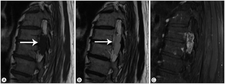
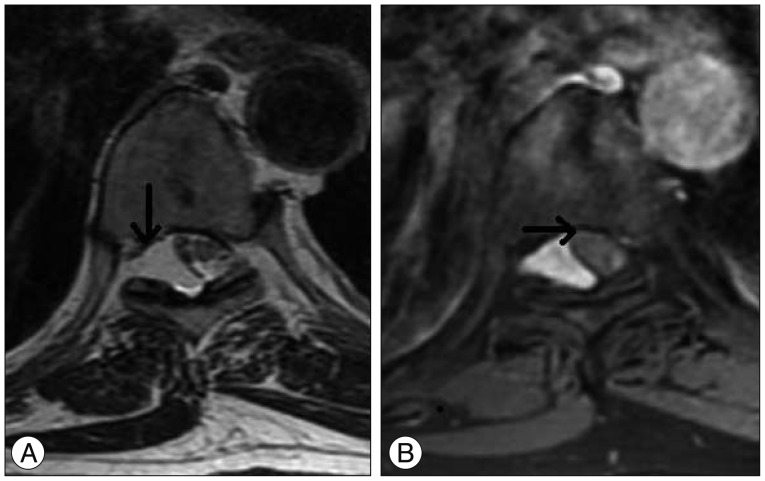
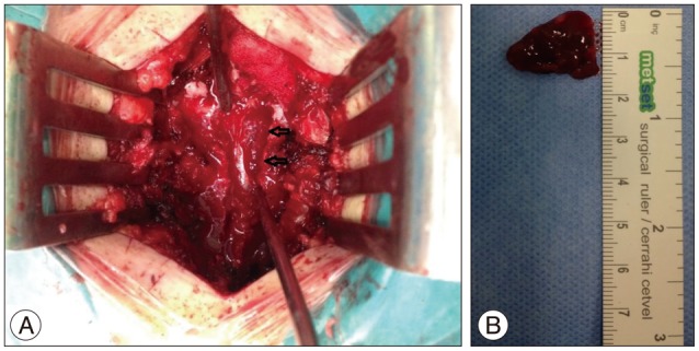
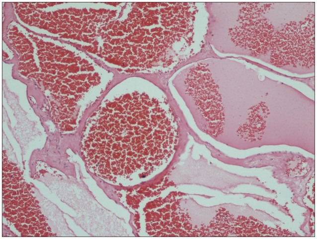
Cavernous hemangiomas were first reported in 1929 by Globus and Doshay, and are defined as benign vascular structures developed between the neural tissues occurring in the central nervous system, consisting of a dilated vascular bed. Cavernous hemangiomas comprise nearly 5-12% of all spinal vascular malformations; however, existence in the epidural space without bone involvement is rare. Only 4% of all cavernous hemangiomas (0.22/1.000.000) are purely epidural cavernous hemangiomas. In this case report, we removed a hemorrhagic thoracic mass presenting with progressive neurological deficits in a 55-year-old male patient. We found this case to be appropriate for presentation due to the rare occurrence of this type of cavernous hemangioma.
Keywords: Cavernous hemangioma; Extraosseous-epidural; Hemangioma; Thoracic vertebrae.
Figures
References
-
- Babu R, Owens TR, Karikari IO, Moreno J, Cummings TJ, Gottfried ON, et al. Spinal cavernous and capillary hemangiomas in adults. Spine (Phila Pa 1976) 2013;38:E423–E430. - PubMed
-
- Feng J, Xu YK, Li L, Yang RM, Ye XH, Zhang N, et al. MRI diagnosis and preoperative evaluation for pure epidural cavernous hemangiomas. Neuroradiology. 2009;51:741–747. - PubMed
-
- Globus JH, Doshay LJ. Venous dilatations and other intraspinal vessel alterations, including true angiomata, with signsand symtoms of cord compression. A report of four cases with areview of the literature. Surg Gynecol Obstet. 1929;48:345–366.
-
- Hegde A, Mohan S, Tan KK, Lim CC. Spinal cavernous malformations : magnetic resonance imaging and associated findings. Singapore Med J. 2012;53:582–586. - PubMed
Publication types
LinkOut - more resources
Full Text Sources
Other Literature Sources
