. 2015 Feb 11;17(2):243-51.
doi: 10.1016/j.chom.2015.01.003.

Herpes simplex virus suppresses necroptosis in human cells

Affiliations

Herpes simplex virus suppresses necroptosis in human cells

Hongyan Guo et al. Cell Host Microbe. .

Abstract

Herpes simplex virus (HSV)-1 and HSV-2 are significant human pathogens causing recurrent disease. During infection, HSV modulates cell death pathways using the large subunit (R1) of ribonucleotide reductase (RR) to suppress apoptosis by binding to and blocking caspase-8. Here, we demonstrate that HSV-1 and HSV-2 R1 proteins (ICP6 and ICP10, respectively) also prevent necroptosis in human cells by inhibiting the interaction between receptor-interacting protein kinase 1 (RIP1) and RIP3, a key step in tumor necrosis factor (TNF)-induced necroptosis. We show that suppression of this cell death pathway requires an N-terminal RIP homotypic interaction motif (RHIM) within R1, acting in concert with the caspase-8-binding domain, which unleashes necroptosis independent of RHIM function. Thus, necroptosis is a human host defense pathway against two important viral pathogens that naturally subvert multiple death pathways via a single evolutionarily conserved gene product.


Figures

Figure 1
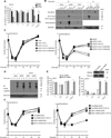
Figure 1. Role of HSV R1 in suppression of virus-induced death
(A) Viability of HSV1 strain KOS or ΔICP6 mutant virus-infected HT-29 cells (MOI=5), or mock-infected, and treated from 2 to 24 hpi with TNF (T; 30 ng/mL), IAP antagonist BV6 (S; 1 µM) and/or caspase inhibitor zVAD (V; 25 µM) alone or in the combinations shown. Viability was determined by measuring ATP levels. Data are normalized to DMSO solvent control (% untreated cells). (B) IB to detect cleaved Casp3 (Cl-Casp3), phosphorylated MLKL (p-MLKL) and β-actin in HT-29 cells infected with KOS or ΔICP6 (MOI=5), or mock-infected, and then treated with T, T+S, T+V, T+S+V or DMSO solvent control (-), from 2 to either 10 or 15 hpi, as indicated. (C) Replication levels of KOS (left) and ΔICP6 (right) virus in HT-29 cells (MOI=5) in DMSO control medium or in medium containing T plus IAP antagonist SMAC007 (S7, 1 µM) (Mandal et al., 2014), T+S7+V or T+S7+V with RIP1 kinase inhibitor GSK’963 (1 µM) or RIP3 kinase inhibitor GSK’840 (3 µM), as shown. Viral titers were determined by plaque assay on Vero cells at the indicated time (hpi). (D) IB to detect viral antigens and β-actin following infection of HT-29 cells with KOS or ΔICP6 virus (MOI=5) in the presence of phosphonoformate (300 µg/ml) and then either left untreated or treated with T+S+V from 2 to 4 or 8 hpi. (E) Viability of HT-29 cells stably expressing nontargeted, scrambled (Sc) shRNA or MLKL-specific shRNA either mock-infected (left) or ΔICP6-infected (right) and then treated with T, T+S, T+V, T+S+V for 24 h. (F) Replication levels of KOS (left) and ΔICP6 (right) virus in HT-29 cells (MOI=5) stably expressing either Sc shRNA or MLKL shRNA in the absence (DMSO control) or presence of T+S7 or T+S7+V as shown. Data with error bars depict mean ± standard deviation (SD); with significance *P <0.05, **P <0.01 and ***P <0.001 indicated above bars. See also Figure S1.
Figure 2
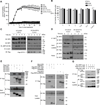
Figure 2. HSV R1 inhibits TNF-induced necroptosis by competing for RHIM-dependent interaction of RIP1 and RIP3
(A) Time course depiction of the accumulation of Sytox Green483 permeable HT-29-EV, HT-29-ICP6 or HT-29-ICP10 cells (images shown in S2A). (B) Viability of HT-29-EV, HT-29-ICP6 or HT-29-ICP10 cells treated for 24 h with T, S, V or the combinations indicated. (C) IB for RIP3, RIP1, phospho-RIP1-S166 and β-actin in cell lysates from HT-29-EV and HT-29-ICP10 cells treated for the indicated times with T+S+V. An asterisk marks slower migrating modified forms of RIP3 or RIP1. (D) Immunoprecipitation/IB (IP/IB) to detect RIP1 interaction with RIP3 in HT-29-EV and HT-29-ICP10 cells. Top panels show IP of cell lysates with anti-RIP1 antibody followed by IB with anti-RIP3 or anti-RIP1 antibody. Middle panels show endogenous RIP3 and RIP1, as well as transduced FLAG-ICP10, in clarified lysates and bottom panels show RIP3 and RIP1 in the pellet fraction. The vertical line shows where lanes from the original gel were brought adjacent. (E) IP/IB to detect ICP6 interaction with RIP kinases. Top panels show lysates of 293T cells transfected with FLAG-ICP6 and Myc-tagged human RIP1, RIP2, RIP3 or RIP4 subjected to IP with anti-Myc antibody followed by IB with anti-FLAG or anti- Myc antibody. (F) IP/IB to detect ICP6 interaction with WT and mutant forms of RIP1. Top panels show lysates of cells transfected with Myc-ICP6 and FLAG-tagged versions of RIP1, RIP1(301–558), RIP1(301–558, mutRHIM), RIP3 or RIP3mutRHIM subjected to IP with anti- FLAG antibody followed by IB with anti-Myc or anti-FLAG antibody. (G) IP/IB demonstrating ICP10 disruption of RIP1 and RIP3 interaction. Top panels show lysates of cells transfected with FLAG-RIP3 with or without Myc-RIP1 in the presence of 0, 0.2, 0.5 or 1.0 µg FLAG-ICP10 subjected to IP with anti-Myc antibody followed by IB with anti-FLAG or anti-Myc antibody. The lower panels depict IB of input cell lysates. Protein molecular size is shown to the right of the lanes. Data with error bars are depict mean ± SD; *P <0.05, **P <0.01, ***P <0.001. See also Figures S2.
Figure 3
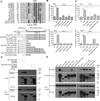
Figure 3. Contribution of RHIM and Casp8-binding domain of R1 to the suppression of TNF-induced necroptosis
(A) The top panel depicts the amino acid sequence of RHIM508 containing regions of human and murine proteins RIP1, RIP3, TRIF and DAI aligned with herpesvirus R1 homologs, MCMV M45, HSV1 ICP6 and HSV2 ICP10. The position of the tetra- Ala mutation (mutRHIM) used to disrupt the RHIM-like motif of ICP6 and ICP10, and used to define the RHIM in other proteins, is indicated below. The bottom panel is a schematic representation and summary of HSV1 R1 mutant proteins. Full-length (aa1-1137) R1 is depicted at the top with an N-terminal (NH2) domain (white) with RHIM (black bar) and C-terminal RR homology domain (gray) as well as the nucleotide binding GXGXXG motif (black circles). FLAG-tagged full length, ICP6mutRHIM, ICP6(Δ1–243) N-terminal deletion, ICP6 N-terminal fragments (1–489, 1–629, 1–835, 1–1106, 1–1116) as well as RHIM mutant form ICP6(1–629, mutRHIM) and central fragment ICP6(244–629) are depicted below. (B) Viability of HT-29 cells stably transduced with EV, ICP6, ICP6(Δ1–243), ICP6mutRHIM, ICP6(1–489), ICP6(1–629) or ICP6(244–629), infected (mock, upper panel; ΔICP6, lower panel) and treated with T+S+V from 2 to 24 hpi. (C) Cell viability of HT-29 cells expressing ICP6(1–835), ICP6(1–1106), ICP6(1–1116) or ICP6(G865/867/870A) infected (mock, upper panel; ΔICP6, lower panel) and treated with T+S+V from 2 to 24 hpi. (D) IP/IB to detect FLAG-ICP6(1–629) or FLAG-ICP6(1–629, mutRHIM) interaction with Myc-RIP1 and Myc-RIP3. IP of cell lysates employed anti-Myc antibody followed by IB with anti-Myc or anti-FLAG antibodies. (E) IP/IB to detect R1 interaction with endogenous Casp8 in HT-29-ICP6, HT-29-ICP10, HT-29-M45 or HT-29 cells expressing FLAG-tagged mutants of ICP6 (Δ1–243, 1–629, 1–835, 1–1106, 1–1116, G865/867/870A and mutRHIM) or ICP10(Δ1–249). IP of cell lysates was with anti-FLAG antibody followed by IB with anti-Casp8 or anti-FLAG antibodies. Protein molecular size is shown to the right of the lanes. Data with error bars depict mean ± SD; *P <0.05, **P <0.01, ***P <0.001. See also Figure S3.
Figure 4
Figure 4. C-terminal interaction of R1 with Casp8 sensitizes cells to necroptosis
(A and C) Viability of HT-29-EV, HT-29-ICP6, HT-29-ICP6mutRHIM and HT-29-ICP6(1–629) cells that were either mock-infected or infected with ΔICP6 or KOS virus (MOI=5) and treated with T or T+S from 2 to 24 hpi. (B and D) IB of p-MLKL, Cl-Casp3, β-actin and FLAG-tagged ICP6 species in HT-29 cells stably expressing EV, ICP6, ICP6mutRHIM and ICP6(1–629). Cell were either mock-infected or infected with KOS or ΔICP6 virus (MOI=5) and then treated with T from 2 to 15 hpi or T+S from 2 to 10 hpi. (E) Viability of HSV1 parental strain F or RHIM mutant virus538 infected HT-29 cells (MOI=10), or mock-infected, and treated from 1 to 24 hpi with T, T+V, T+GSK’963, or T+GSK’840. (F) IB to detect cleaved p-MLKL, Cl-Casp3, and β-actin in HT-29 cells infected with HSV1(F strain) or HSV1mutRHIM (MOI=1.5), or mock-infected, and then treated with T from 1 to 20 hpi. (G and H) Replication levels of HSV1 and HSV1mutRHIM virus in HT-29 cells (MOI=5) in DMSO control medium or in medium containing T, T+V or T+ V in the presence or absence RIP1 kinase inhibitor GSK’963 (1 µM) or RIP3 kinase inhibitor GSK’840 (3 µM) as shown. Viral titers were determined by plaque assay on Vero cells at the indicated time (hpi). Data with error bars depict mean ± SD; *P <0.05, **P <0.01, ***P <0.001. See also Figure S4.

Comment in

  • HSV cheats the executioner.
    Mossman KL, Weller SK. Mossman KL, et al. Cell Host Microbe. 2015 Feb 11;17(2):148-51. doi: 10.1016/j.chom.2015.01.013. Cell Host Microbe. 2015. PMID: 25674981

Similar articles

Cited by

References

    1. Berger AK, Danthi P. Reovirus activates a caspase-independent cell death pathway. mBio. 2013;4:e00178–e00113. - PMC - PubMed
    1. Bousfiha A, Picard C, Boisson-Dupuis S, Zhang SY, Bustamante J, Puel A, Jouanguy E, Ailal F, El-Baghdadi J, Abel L, et al. Primary immunodeficiencies of protective immunity to primary infections. Clin Immunol. 2010;135:204–209. - PubMed
    1. Brune W, Menard C, Heesemann J, Koszinowski UH. A ribonucleotide reductase homolog of cytomegalovirus and endothelial cell tropism. Science. 2001;291:303–305. - PubMed
    1. Chabaud S, Sasseville AM, Elahi SM, Caron A, Dufour F, Massie B, Langelier Y. The ribonucleotide reductase domain of the R1 subunit of herpes simplex virus type 2 ribonucleotide reductase is essential for R1 antiapoptotic function. J Gen Virol. 2007;88:384–394. - PubMed
    1. Chan FK, Shisler J, Bixby JG, Felices M, Zheng L, Appel M, Orenstein J, Moss B, Lenardo MJ. A role for tumor necrosis factor receptor-2 and receptor-interacting protein in programmed necrosis and antiviral responses. J Biol Chem. 2003;278:51613–51621. - PubMed

Publication types

MeSH terms